MEN'S HEALTH I

CANCER

Advances in surgical treatment for prostate carcinoma

A new group of focused treatments aim to surgically treat disease without removal of the gland

Dr Ray McDermott, Consultant Medical Oncologist, Tallaght Hospital, Dublin, Dr John Donnellan, Radiology SpR, Tallaght Hospital, Dublin, Mr Shane Considine, Urology Registrar, St James’s Hospital, Dublin, Mr Rustom Manecksha, Consultant Urologist, St James's Hospital, Dublin, Dr Emmet Jordan, Oncology Specialist Registrar, Tallaght Hospital, Dublin and Dr John Feeney, Consultant Radiologist, Tallaght Hospital, Dublin

February 4, 2015

Article
Similar articles
  • Surgery plays a key role in the management of prostate cancer, particularly in organ-confined disease, but also in some locally advanced disease and in post-radiotherapy relapse. 

    The standard approach to surgical management of prostate cancer is radical retropubic prostatectomy (RRP), which involves removal of the entire prostate gland and seminal vesicles, and anastomosis of the urethra to the bladder neck. The basic principles of this surgery have remained largely unchanged for many years, however, there have been certain refinements undertaken to make the surgery less morbid and less invasive. We have also seen the emergence of a new group of ‘focused treatments’ that aim to surgically treat disease without removal of the gland.

    Radical prostatectomy

    In prostate cancer surgery, the priorities for the surgeon are removal of all malignant tissue, preservation of urinary continence and maintenance of erectile function (where possible), in that order. As such, these endpoints are generally used for comparison of novel surgical techniques.

    Minimally invasive surgery for prostate cancer has emerged as a viable alternative to the traditional open operation. This can be performed either by standard laparoscopic means or by a robot-assisted technique. The latter is fast becoming the surgical option of choice in the US. In Ireland, there are currently two da Vinci robots in urological operation and several surgeons offering robotic prostatectomy, while laparoscopic prostatectomy is performed by a number of surgeons also. 

    Despite being relatively new advances, both laparoscopic and robotic prostatectomy have been shown to result in equivalent oncological outcomes as open surgery. A systematic review by Nevara et al1 showed no significant difference in rates of positive surgical margins or progression-free survival between the three techniques. However, they do note that follow-up beyond five years from robot-assisted surgery was limited to a small number of papers. 

    Recently, five-year biochemical recurrence rates of 14% and mortality rates of 1% have been reported with robot-assisted surgery,2 while 10-year progression-free survival of 78.1-97.2% (depending on stage) has been shown with the laparoscopic technique.3

    In terms of morbidity, lower rates of blood loss and transfusion are reported with robotic surgery compared to RRP,4 as well as a significantly lower incidence of urinary incontinence compared to laparoscopic surgery and RRP.5 Interestingly, while robotic surgery was associated with lower rates of impotence than RRP, no superiority was demonstrated over laparoscopic surgery.6

    While there is general agreement that robotic prostatectomy is a valuable surgical technique, the Pasadena Consensus Panel concluded that studies with longer follow-up are required to fully define its role and efficacy.7

    Focused treatments

    With the increase in opportunistic screening with prostate-specific antigen (PSA) in recent years, we are now diagnosing more and more early-stage, low-grade prostate cancers. As such, there is a drive for the development of low-morbidity definitive treatment strategies. Much research in recent times has concentrated on focused surgical treatments for prostate cancer, in which the pathologic areas of the gland are treated with a minimum of injury to the normal prostate tissue. 

    Two candidate therapies have emerged to date, namely cryotherapy and high intensity focused ultrasound (HIFU), while vascular-targeted photodynamic therapy remains in the experimental stages. Given that it is targeted at the areas of malignancy specifically, it is imperative that accurate localisation of disease is achieved prior to treatment. This mandates either transperineal template biopsies (which can achieve a sensitivity of up to 90%8) or, if unavailable, new multiparametric MRI and transrectal ultrasound (TRUS) biopsy.9

    One of the major difficulties with research in this field is the lack of a consistent definition for biochemical recurrence, with different groups utilising different thresholds. Since not all prostate tissue is destroyed, we would expect there to be a non-zero PSA level following treatment, but how to interpret this level remains unclear.

    Cryosurgery

    Cryosurgery as a discipline has been practised for many years and involves the sequential freezing and thawing of the prostate gland to cause tissue damage and cell death. The patient is placed in the lateral position, under either general or regional anaesthesia, and a number of cryoprobes (up to 30) are placed transperineally into the prostate gland under TRUS guidance. Argon gas is passed through these probes resulting in rapid cooling of the prostate tissues. A urethral warmer is utilised and all probes must be placed at least 8mm from the urethra to prevent tissue sloughing and urethral injury. 

    Thermosensors are placed at the external sphincter, at the apex of the gland and along Denonvilliers’ fascia to ensure that the tissues reach a temperature of -40ºC, and are maintained at this level for three minutes. Following this, the argon gas is exchanged for helium resulting in thawing of the probes. 

    Two freeze-thaw cycles have been shown to result in better oncological outcomes than a single cycle10 as well as tissue destruction at a temperature of -41ºC, rather than the -62ºC needed with a single cycle.11

    The European Association of Urology (EAU) identifies this as a potentially useful modality in low-to-intermediate-risk prostate cancer in those with prostate size of less than 40g.9

    Cryosurgery has been used both as a primary therapeutic modality in prostate cancer and as a salvage treatment for relapse following radiotherapy.12 In the primary setting, five-year actuarial biochemical recurrence-free survival ranged from 36-61% (low-risk to high-risk disease) and 45-76% depending on whether 0.5 or 1.0ng/ml PSA was used as a threshold.13 The overall rate of positive prostate biopsy after therapy was 18%.13

    As expected, the results for salvage cryotherapy for patients with recurrence after radiotherapy are worse, with Williams et al reporting a five-year survival of 39%.14

    The major complication associated with cryosurgery is erectile dysfunction, with rates of 93% reported,13 improving to 51% at four years with formal penile rehabilitation.15 Sexual function at three years post-treatment was shown to be superior to patients following external beam radiotherapy. Ahmed et al demonstrated incontinence rates of 5% following primary and 10% following salvage cryosurgery.16 Other complications include tissue sloughing, pelvic pain, urinary retention and, rarely, fistula formation.

    High intensity focused ultrasound

    High intensity focused ultrasound (HIFU) is the other major modality of focal treatment for prostate cancer under investigation. It is also performed under general anaesthesia by a transperineal approach. High intensity ultrasound waves are focused onto the area of abnormality in the gland, resulting in heating of the tissues and coagulative necrosis. Unfortunately, like cryotherapy, despite having been in use for several years, the quality of evidence for this procedure is lacking, with no randomised controlled trials available. 

    A recent systematic review of 31 uncontrolled trials reported negative post-treatment biopsy rates of 35-95%, and five-year survival of 61.2-95%.17 Blana et al have shown recurrence rates post-HIFU to be related to the level of the PSA nadir, with those achieving lowest post-op PSA levels having the best outcome. As expected, the prognosis associated with this modality is related to the stage at time of diagnosis, with a biochemical disease-free rate at five years post-treatment varying from 74% for T1c disease to 33% for T3.18

    Complications encountered with HIFU include urinary retention, erectile dysfunction, urinary incontinence, UTIs and, rarely, recto-urethral fistula. Cordiero et al report that the incidence of obstructive complications can be reduced by TURP, either at the time of HIFU or postoperatively.17

    Vascular-targeted photodynamic therapy

    The technique of vascular-targeted photodynamic therapy, while currently still in the experimental stage of development, may in future be added to the range of focal therapies available. This approach aims to destroy cancer cells by the activation of a systemically administered photodynamic agent by local application of LASER light under TRUS guidance,19 allowing targeting of therapy and minimisation of complications.

    Despite some encouraging results in the field of focal therapy, both the EAU9 and NICE20 advise that these modalities (in general) remain within the field of clinical trials until longer-term follow-up is available. However, the American Urological Association suggests,21 and the EUA concedes, that cryotherapy may be considered in selected cases, particularly in those unfit for radical surgery or with limited life expectancy. 

    Radiology

    The advent of early detection methods for prostate cancer has led to a requirement for more definitive diagnostic modalities. The traditional imaging sequences are being pressed to offer greater detail and definition so as to have minimally invasive, patient-centred, targeted therapies available to more patients diagnosed with prostate cancer. The latest developments have been focused largely on magnetic resonance imaging (MRI) as well as therapeutic nuclear medicine options.

    Multiparametric MRI

    Multiparametric MRI is a term that has been adopted to incorporate the multitude of MR-based imaging techniques currently being employed in the detection of prostate cancer, among others. The term is usually employed to describe the use of MR spectroscopy, dynamic contrast-enhanced MR and diffusion-weighted imaging, or variations thereof. 

    MR spectroscopy 

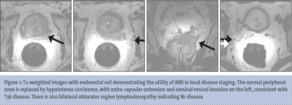
    MR spectroscopic imaging (MRSI) generates information about the relative concentrations of specific metabolites within tissues and is commonly performed in 3D imaging of the brain.22 When used for prostate imaging, an endorectal coil is recommended for 3 tesla (3T) magnets and required for 1.5T magnets.23 T2-weighted images are generated (see Figure 1) and are overlaid then by spectral tracings generated by MRSI. MRSI analyses a defined area for specific molecules such as hydrogen ions or protons. 

     (click to enlarge)

    Knowing the structure of the metabolite of interest allows the relative concentrations of different metabolites to be plotted on a graph versus normal prostatic tissue, thus showing the relative peaks and troughs of the different compounds of interest within the interrogated voxel. In prostate cancer, citrate and choline are the metabolites of most interest.24 Choline has been shown to be present in prostate carcinoma at higher concentrations than in the normal prostate tissue. Citrate is reduced in carcinoma compared with normal prostatic tissue. It is the ratio of choline to citrate that is used to assess a given voxel for likelihood of prostate carcinoma. 

    While MRSI is being heavily developed, particularly in large research centres, it is time consuming and often challenging and as such is not routinely performed in most institutions. Currently, best evidence shows T2-weighted imaging at 1.5T is as efficient as MRSI with T2WI in tumour localisation and is listed as optional, not routine, by the recent guidelines from the European Society of Urogenital Radiology (ESUR).22 However, as it evolves, MRSI is likely to play an increasing role in detection of early prostate cancer.

    Diffusion-weighted imaging

    Diffusion-weighted imaging (DWI) provides information about tissue microstructure and specifically the ability of water molecules to diffuse through that region. Its utility in stroke imaging has been widely published. The signal generated in a diffusion sequence is directly related to the ability of the water molecules to move freely within the space being interrogated whereby restricted molecules will appear bright on the diffusion sequence. By changing the diffusion gradient applied, the relative freedom of the molecules can be ascertained and used to generate an apparent diffusion coefficient (ADC) map which does not suffer from the confounding T2-weighting that native DWI does. 

    The high cellular density present in prostate cancers, and in many tumours, leads to restricted water diffusion, which is detectable on DWI.23 The latest meta-analysis by Tan et al25 has shown a sensitivity and specificity of 69% and 89%, respectively, for DWI alone in detection of prostate cancer. Current research is showing an inverse correlation between ADC values derived from DWI images and Gleason scores of prostate cancer on biopsy.26 It is hoped that ultimately this will spare some patients from needing to undergo a TRUS biopsy, which can have variable sensitivity, particularly for transitional zone tumours,27 thereby saving them from an invasive and often poorly tolerated procedure (see Figure 2).

     (click to enlarge)

    Dynamic contrast-enhanced MRI

    Angiogenesis in tumour cells can potentially lead to variations in perfusion, vascular density and turbulent blood flow, delineating it from the normal underlying tissue.28 There are many different ways to perform dynamic contrast-enhanced MRI (DCEMRI), however the majority are based on a spoiled gradient echo sequence, T1-weighted, which is run before the rapid infusion of a gadolinium chelate compound, and then repetitively re-run as the contrast perfuses to generate diagnostic images. 

    The different methods of DCEMRI have led to an explosion in ways to produce the images, as well as differences in interpretation protocols, ranging from qualitative analyses based on colour-coded maps, through semi-quantitative to fully quantitative, which sets out to measure the actual concentration of gadolinium in the tissues of interest through the utility of pharmacokinetic modelling.23 There is research published showing the advantages and disadvantages of each method, however the ESUR has recently published guidelines recommending the semi-quantitative approach using analysis of kinetic curves for DCEMRI.22

    Recently, research has shown that the fully quantitative approach can also allow for the monitoring of treatment response, which would be of significant benefit.29 Ultimately, however, the choice of DCEMRI technique and analysis is heavily dependent on the hardware and software configuration available on an institution-by-institution basis.

    Radium-223

    A novel treatment option for castrate-resistant metastatic prostate cancer has recently become available in the US in the form of radium-223, the first ever licensed therapeutic alpha emitter. This treatment is currently undergoing extensive clinical trials and is expected to be available to suitable patients shortly. While beta-particle emitters (strontium-89, samarium-153) have a well-established role in bone pain palliation, radium-223 is the first radiotherapeutic agent to demonstrate an additional survival benefit.

    More than 90% of patients with castrate-resistant metastatic prostate cancer have radiographic evidence of bony disease.30,31 In contrast to many other cancers, death from prostate cancer is often due to bone disease and complications,32 which is why current treatments such as bisphosphonates, denosumab and existing radioisotope treatments have primarily focused on pain relief and deference of bony events.33,34,35,36,37,38,39

    A study evaluating denosumab versus zoledronic acid in castrate-resistant prostate cancer assessed 1,904 patients who were randomised, 950 assigned to denosumab and 951 assigned to receive zoledronic acid. Median time to first on-study skeletal-related event was 20.7 months (95% CI, 18.8-24.9) with denosumab compared with 17.1 months (15.0-19.4) with zoledronic acid (HR 0.82, 95% CI, 0.71-0.95; p = 0.0002 for non-inferiority; p = 0.008 for superiority). Rates of hypocalcaemia occurred more frequently in the denosumab group (121 [13%]) than in the zoledronic acid group (55 [6%]; p < 0.0001). Osteonecrosis of the jaw occurred infrequently (22 [2%] in the denosumab group versus 12 [1%] in the zoledronic acid subgroup; p = 0.09).37 Supplementation of at least 500mg calcium and 400 IU vitamin D daily is required in all patients on denosumab to reduce the risk of hypocalcaemia.

    Radium-223 is a radioisotope that emits high-energy alpha particles. Alpha particles are in essence radioactive helium nuclei that have a high linear energy transfer (LET) value, meaning that they deposit a lot of energy over a short distance, somewhere in the region of 100µm (half the thickness of a human hair).40 This means that as a result of the short distance that alpha particles can travel, the toxic effects on adjacent tissue, and potentially bone marrow, are minimised.41,42

    As a bone-seeking calcium mimetic, radium-223 is bound especially within the microenvironment of osteoblastic or sclerotic metastases.41,43 The high-energy alpha-particle radiation induces formation of double-stranded DNA breaks resulting in a potent, localised cytotoxic effect. A phase III, randomised, double-blind, placebo-controlled study assessed 921 patients who were randomised to receive six injections of radium-223 (dose of 50kBq per kilogram of body weight intravenously) or matching placebo; one injection was administered every four weeks. Updated analysis confirmed the survival benefit of radium-223 (median 14.9 months versus 11.3 months; HR, 0.70; 95% CI, 0.58 to 0.83; p < 0.001.40 A low myelosuppression rate was associated with its use.

    In summary, radium-223 has been shown to have a favourable safety profile, with minimal toxicity, to reduce pain, improve disease-related markers and improve survival with a 30% reduction in risk of death, for patients with mCRPC,40 however clinical trials are still ongoing.

    Radiotherapy in prostate cancer

    Primary external beam radiation

    External beam radiotherapy (EBRT) is a well established treatment modality for localised prostate carcinoma. It is a standard approach for patients with intermediate and high-risk prostate cancer in combination with androgen deprivation therapy (ADT). The addition of ADT has been shown to improve both disease-free and overall survival.44,45 Doses of 75.6-79.2Gy can be used for low-risk prostate cancers, while doses of 81Gy can be used for intermediate and high-risk disease. There are very few contraindications for EBRT compared to prostate brachytherapy. 

    EBRT can be offered to men for whom general or spinal anaesthetic is contraindicated, patients with severe comorbidities, a large prostate size (> 70g) or high-risk disease. EBRT is generally associated with fewer urinary side-effects but higher rectal toxicity than that seen with brachytherapy.46 EBRT is usually delivered over a period of seven to eight weeks of daily fractions. Combination of EBRT and brachytherapy is considered to be more effective than EBRT alone in some intermediate-risk patients and in high-risk patients.

    Prostate brachytherapy

    Prostate brachytherapy (PB) has gained acceptance as a treatment option in prostate cancer in recent years. Brachytherapy refers to placement of radioactive sources inside or adjacent to a cancerous tumour. There are two different forms of prostate brachytherapy.

    Low-dose-rate (LDR) brachytherapy: This is where radioactive seeds are implanted permanently into the prostate. This allows delivery of adequate doses of radiation to the cancer while avoiding toxicity to bladder and rectum. Low-dose-rate PB is a day-case procedure that takes about one hour. It is performed under general or spinal anaesthesia (occasionally local). Patients have minimal recovery time post-procedure so there is minimal effect on activities of daily living. Between 70 and 150 radioactive seeds are implanted into the prostate through the perineum, using 20-30 needles, each carrying two to seven seeds.46

    Many men experience some moderate irritative and obstructive urinary symptoms lasting several months. More than 90% have minimal or no urinary symptoms in the long term.47,48,49 In approximately 5-10% of patients urinary retention may occur that can require catheter insertion. These episodes of urinary retention usually resolve within a few days. Self-limiting rectal irritation affects 20-30% of patients and is mild in nature. Rectal bleeding can occur in 2-7% of patients.50 Serious rectal injury requiring a major intervention surgically such as colostomy is very rare (fewer than one in every 500-1,000 patients). Erectile dysfunction rates after brachytherapy are favourable. 

    Patients with low-risk disease (clinical stage ≤ T2a, PSA level ≤ 10.0ng/mL or Gleason scores ≤ 6) are eligible for brachytherapy. For patients with intermediate-risk disease PB may be used in combination with EBRT.46 Patients with small prostates, symptoms of urinary retention or a history of TURP procedure are not ideal candidates for PB as complication rates are increased. Post-implant dosimetry should be performed post-procedure to confirm satisfactory implantation. The monotherapy dose is 145Gy for iodine-125 and 125Gy for palladium-103.

    High-dose-rate (HDR) brachytherapy: This is where treatment is administered over approximately 10 minutes through temporary catheters that contain the radioactive sources. High-dose-rate brachytherapy is usually used in combination with EBRT for intermediate or high-risk disease. Addition of ADT to brachytherapy and EBRT has been used in patients with a high risk of recurrence. This tri-modality treatment approach had nine-year progression-free and disease-specific-free survival of 87% and 91%, respectively.50,51 External beam radiation therapy is commonly delivered over a period of seven to eight weeks of daily treatments.40 Vargus et al reported a lower risk of erectile dysfunction with HDR than LDR.52 Two groups reported a lower rate of urinary frequency, urgency and rectal pain with HDR than LDR.53,54

    Palliative radiation can be used for symptomatic bone metastases. A short course of 8Gy for one day is as effective as 30Gy in 10 fractions and is also less costly. Radium-223, a radioactive alpha particle emitting agent, has been evaluated in metastatic castrate-resistant prostate cancer with symptomatic bone metastases and is discussed elsewhere in this article. 

    Conclusion

    Since the incidence of prostate cancer is highest in elderly men, an important consideration must be given to life expectancy, comorbidities, current functional status and the ability of a patient to tolerate potential toxicities associated with these therapies when deciding treatment. Because elderly patients also may benefit from chemotherapies to the same degree as younger patients, physicians must appropriately assess patients and derive appropriate treatment strategies to optimise patient outcomes so that they can get the most benefit from active agents.7

    Identification of biomarkers may help define the optimum treatment paradigm in the changing landscape of prostate carcinoma and help to identify patient subgroups that are more likely to benefit from each of these active agents.8

    Further research which will help to assess and identify the best sequencing of these agents, their use alone or as part of combination strategies. Certainly this is a welcome conundrum in the treatment of prostate cancer. Since the docetaxel era in 2004, indisputably progress has been made in this disease. It is now at the stage where we must learn how to piece it all together to make the most use of these active agents in prostate cancer. Exciting times lie ahead.

    References 

    1. Novara G et al. Systematic review and meta-analysis of studies reporting oncologic outcome after robot-assisted radical prostatectomy. Eur Urol 2012; 62(3): 382-404.
    2. Liss MA et al. Robot-assisted radical prostatectomy: 5-year oncological and biochemical outcomes. J Urol 2012; 188(6): 2205-10.
    3. Hruza M et al. Long-term oncological outcomes after laparoscopic radical prostatectomy. BJU Int 2013; 111(2): 271-80.
    4. Novara G et al. Systematic review and meta-analysis of perioperative outcomes and complications after robot-assisted radical prostatectomy. Eur Urol 2012; 62(3): 431-52.
    5. Ficarra V et al. Systematic review and meta-analysis of studies reporting urinary continence recovery after robot-assisted radical prostatectomy. Eur Urol 2012; 62(3): 405-17.
    6. Ficarra V et al. Systematic review and meta-analysis of studies reporting potency rates after robot-assisted radical prostatectomy. Eur Urol 2012; 62(3): 418-30.
    7. Montorsi F et al. Best practices in robot-assisted radical prostatectomy: recommendations of the Pasadena Consensus Panel. Eur Urol 2012; 62(3): 368-81.
    8. Crawford ED et al. Clinical staging of prostate cancer: a computer-simulated study of transperineal prostate biopsy. BJU Int 2005; 96(7): 999-1004.
    9. Heidenreich A et al. EAU Guidelines on Prostate Cancer. Part 1: Screening, Diagnosis, and Local Treatment with Curative Intent-Update 2013. Eur Urol 2014; 65(1): 124-37.
    10. Shinohara K et al. Cryosurgical ablation of prostate cancer: patterns of cancer recurrence. J Urol 1997; 158(6): 2206-9; discussion 2209-10.
    11. Larson TR et al. In vivo interstitial temperature mapping of the human prostate during cryosurgery with correlation to histopathologic outcomes. Urology 2000; 55(4): 547-52.
    12. Spiess PE, Given RW, Jones JS. Achieving the ‘bifecta’ using salvage cryotherapy for locally recurrent prostate cancer: analysis of the Cryo On-Line Data (COLD) registry data. BJU Int 2012; 110(2): 217-20.
    13. Long JP et al. Five-year retrospective, multi-institutional pooled analysis of cancer-related outcomes after cryosurgical ablation of the prostate. Urology 2001; 57(3): 518-23.
    14. Williams AK et al. Disease-free survival following salvage cryotherapy for biopsy-proven radio-recurrent prostate cancer. Eur Urol 2011; 60(3): 405-10.
    15. Ellis DS. Cryosurgery as primary treatment for localized prostate cancer: a community hospital experience. Urology,2002; 60(2 Suppl 1): 34-9.
    16. Ahmed S, Davies J. Managing the complications of prostate cryosurgery. BJU Int 2005; 95(4): 480-1.
    17. Cordeiro ER et al. High-intensity focused ultrasound (HIFU) for definitive treatment of prostate cancer. BJU Int 2012; 110(9): 1228-42.
    18. Uchida T et al. Transrectal high-intensity focused ultrasound for the treatment of localized prostate cancer: eight-year experience. Int J Urol 2009; 16(11): 881-6.
    19. Marberger M et al. New treatments for localized prostate cancer. Urology 2008; 72(6 Suppl): S36-43.
    20. Prostate Cancer: Diagnosis and Treatment. NICE Guidelines 2008 [cited 2014 06/01/2014]; Available from: http://www.nice.org.uk/nicemedia/live/11924/39687/39687.pdf.
    21. Babaian RJ et al. Best Practice Statement on Cryosurgery for the treatment of localized Prostate Cancer. American Urological Association Guidelines 2008 06/01/2014]; AUA Guideline]. Available from: http://www.auanet.org/education/guidelines/cryosurgery.cfm - 14.
    22. Barentsz JO et al. ESUR prostate MR guidelines 2012. European Radiol 2012; 22(4): 746-757.
    23. Gupta RT et al., The State of Prostate MRI in 2013. Oncology 2013; 27(4): 1.
    24. Weinreb JC et l. Prostate Cancer: Sextant Localization at MR Imaging and MR Spectroscopic Imaging before Prostatectomy – Results of ACRIN Prospective Multi-institutional Clinicopathologic Study1. Radiology 2009; 251(1): 122-133.
    25. Tan CH et al. Diffusion-weighted MRI in the detection of prostate cancer: meta-analysis. Am J Roentgenology 2012; 199(4): 822-9.
    26. Woodfield CA et al. Diffusion-weighted MRI of peripheral zone prostate cancer: comparison of tumour apparent diffusion coefficient with Gleason score and percentage of tumour on core biopsy. Am J Roentgenology 2010; 194(4): 316-22.
    27. Collins GN et al. Multiple transrectal ultrasound-guided prostatic biopsies – true morbidity and patient acceptance. Br J Urol 1994; 71(4): 460-464.
    28. Verma S et al. Overview of dynamic contrast-enhanced MRI in prostate cancer diagnosis and management. Am J Roentgenology 2012; 198(6): 1277-1288.
    29. Low RN, Fuller DB, Muradyan N. Dynamic gadolinium-enhanced perfusion MRI of prostate cancer: assessment of response to hypofractionated robotic stereotactic body radiation therapy. Am J Roentgenology 2011; 197(4): 907-915.
    30. Tannock IF et al. Docetaxel plus prednisone or mitoxantrone plus prednisone for advanced prostate cancer. NEJM 2004; 351(15): 1502-1512.
    31. Roodman GD. Mechanisms of bone metastasis. NEJM 2004; 350(16): 1655-1664.
    32. Lange PH, Vessella RL. Mechanisms, hypotheses and questions regarding prostate cancer micrometastases to bone. Cancer Metastasis Rev 1998; 17(4): 331-336.
    33. Lipton A. Implications of bone metastases and the benefits of bone-targeted therapy. Seminars in Oncology. Elsevier 2010. 
    34. Adami S. Bisphosphonates in prostate carcinoma. Cancer 1997; 80(S8): 1674-1679.
    35. Silberstein EB. Systemic radiopharmaceutical therapy of painful osteoblastic metastases. Seminars in Radiation Oncology. Elsevier 2000.
    36. Fizazi K et al. Randomized phase II trial of denosumab in patients with bone metastases from prostate cancer, breast cancer, or other neoplasms after intravenous bisphosphonates. J Clin Oncol 2009; 27(10): 1564-1571.
    37. Fizazi K et al. Denosumab versus zoledronic acid for treatment of bone metastases in men with castration-resistant prostate cancer: a randomised, double-blind study. The Lancet 2011; 377(9768): 813-822.
    38. Finlay IG, Mason MD, Shelley M. Radioisotopes for the palliation of metastatic bone cancer: a systematic review. The Lancet Oncol 2005; 6(6):  392-400.
    39. Sartor O, Goeckeler W, Bruland O. Stromal targeted therapy in bone metastatic prostate cancer: promise delivered. Asian J Andrology 2011; 13(6): 783-784.
    40. Parker C et al. Alpha emitter radium-223 and survival in metastatic prostate cancer. NEJM 2013; 369(3): 213-223.
    41. Henriksen G et al. Targeting of osseous sites with alpha-emitting 223Ra: comparison with the beta-emitter 89Sr in mice. J Nuclear Med 2003; 44(2): 252-259.
    42. Kerr C. 223Ra targets skeletal metastases and spares normal tissue. The Lancet Oncol 2002; 3(8): 453.
    43. Henriksen G et al. Significant antitumour effect from bone-seeking, alpha-particle-emitting 223Ra demonstrated in an experimental skeletal metastases model. Can Res 2002; 62(11): 3120-3125.
    44. Bolla M et al. External irradiation with or without long-term androgen suppression for prostate cancer with high metastatic risk: 10-year results of an EORTC randomised study. The Lancet Oncol 2010; 11(11): 1066-1073.
    45. Pilepich MV et al. Androgen suppression adjuvant to definitive radiotherapy in prostate carcinoma – long-term results of phase III RTOG 85-31. Int J Rad Oncol* Biol* Physics 2005; 61(5): 1285-1290.
    46. Keyes M et al. Treatment options for localized prostate cancer. Can Fam Phys 2013; 59(12): 1269-1274.
    47. Pickles T, Keyes M, Morris WJ. Brachytherapy or conformal external radiotherapy for prostate cancer: a single-institution matched-pair analysis. Int J Rad Oncol* Biol* Physics 2010; 76(1): 43-49.
    48. Keyes M et al. Predictive factors for acute and late urinary toxicity after permanent prostate brachytherapy: long-term outcome in 712 consecutive patients. Int J Rad Oncol* Biol* Physics 2009; 73(4): 1023-1032.
    49. Crook JM et al. Comparison of health-related quality of life 5 years after SPIRIT: Surgical Prostatectomy Versus Interstitial Radiation Intervention Trial. J Clin Oncol 2011; 29(4): 362-368.
    50. Martínez-Monge R et al. External-Beam Radiation Therapy and High–Dose Rate Brachytherapy Combined With Long-Term Androgen Deprivation Therapy in High and Very High Prostate Cancer: Preliminary Data on Clinical Outcome. Int J Rad Oncol * Biol * Physics 2012; 82(3): e469-e476.
    51. Bittner N et al. Long-term outcome for very high-risk prostate cancer treated primarily with a triple modality approach to include permanent interstitial brachytherapy. Brachytherapy 2012; 11(4): 250-255.
    52. Vargas C et al. A new model using number of needles and androgen deprivation to predict chronic urinary toxicity for high or low dose rate prostate brachytherapy. J Urology, 2005; 174(3): 882-887.
    53. Hoskin P. High dose rate brachytherapy for prostate cancer. Cancer/Radiotherapie 2008; 12(6): 512-514.
    54. Grills IS, et al. High dose rate brachytherapy as prostate cancer monotherapy reduces toxicity compared to low dose rate palladium seeds. J Urology 2004; 171(3): 1098-1104.
    © Medmedia Publications/Cancer Professional 2015