The aim of osteoporosis treatment is prevention of fractures
Dr John Carey, Rheumatology Consultant, Rheumatology Department, Galway University Hospital, Galway and Dr Orla Ni Mhuircheartaigh, Senior House Officer, Rheumatology Department, Galway University Hospital, Galway
Osteoporosis is the most common disease of the skeleton, resulting in compromised bone strength and a subsequent increased risk of fractures.1 The prevalence is rising as longevity increases. Current estimates suggest that approximately 300,000 Irish citizens have osteoporosis, a figure expected to double over the next 20 years.2 This condition is commonly seen in primary care3 where it can usually be diagnosed and managed successfully. Fractures are the most important clinical manifestation of osteoporosis, which can have significant morbidity, increased mortality and costs the state nearly e400 million annually. Fractures are preventable by diagnosing osteoporosis before a fracture occurs, or identifying those who are at risk, and instituting effective preventative measures.
Pathogenesis
Bone strength reflects its composition, mass and structure. Bone mineral density (BMD) is responsible for 70% of bone strength,4 while the remaining 30% relates to the organisation of these minerals along with the distribution of other bone components such as the protein content.5 BMD is 70-80% genetic; the remainder depends on lifestyle choices, diseases and medications.6
Peak bone mass is achieved in early adulthood, depending on the skeletal site and individual. Later bone loss occurs at different sites and rates, most notably at the spine and hips. With normal life expectancy, women lose 40-50% of their bone mass and men 30-40% during the remainder of their lives.6 Genes and hormonal factors have a large influence on bone health, but nutrition, lifestyle, illness and medications are also key determinants of this process.7-9
Risk factors
Risk factors for osteoporosis can be divided into modifiable and non-modifiable. While both are important for identifying the population or individuals at risk, modifiable risk factors have the potential to be rectified, optimising management.
Non-modifiable risk factors:
Genetics: up to 80 different genes have been identified as affecting BMD.10 Limited insights have been gained to date on the importance of each on skeletal fragility
Gender: women account for almost three times the number of osteoporotic fractures as men. This is because men have greater bone acquisition during puberty, reduced bone loss, and probably most importantly, males do not experience menopause or oestrogen loss11
Age: Bone loss occurs with ageing. The older the population, the higher the risk of fracture12
Ethnicity: Rates of osteoporosis are higher in Asian and European populations, but these figures are less durable when adjusted for variations in body size.13
Modifiable risk factors
Numerous modifiable risk factors exist, related to the supply of nutrients to bone, and stimulation of skeletal processes.
Exercise: Activity levels during childhood play an important role in the laying down of bone. Muscles attached to bone act as a stimulus for the osteoblasts to produce bone. Weight-bearing forces help arrange bone in a manner capable of resisting impact. Bone is dynamic and decreased activity levels consequently increase bone resorption8
Nutrition: A healthy balanced diet is essential for skeletal growth and homeostasis. Deficiencies, particularly of protein, calcium and vitamin D result in impaired skeletal composition and defective repair.8,9
A growing list of diseases and medications are known to affect bone (see Table 1).8,9,14,15 While the exact pathobiologic pathways may vary, the net result is impaired bone strength, decreased balance and muscle strength, with consequent increase in fracture risk.
Table 1(click to enlarge)
Diagnosis
Osteoporosis can be diagnosed in three ways: by the presence of a fragility fracture; by bone densitometry of the hip, spine and occasionally radius; or by histomorphometric analysis of a tetracycline-labelled bone biopsy. The latter is seldom used, so only the first two will be discussed here.16
A fragility fracture is a fracture that occurs following, or in the absence of, low impact trauma. Healthy adult bone should be able to withstand the impact from a fall from a standing position, and fractures occurring from forces below this threshold are considered fragility fractures.
In severe osteoporosis, minimum movements such as coughing or laughing, or a slip and fall, can induce a fragility fracture. The most commonly affected sights include the vertebrae (45%), hip (20%) and wrist and forearm (18%). Fragility fractures are diagnosed based on careful history, examination and radiological evidence of a fracture. Only one-third of vertebral fractures present with pain, so doctors need to be mindful of this when assessing older patients. Suggestive physical examination features include height loss (> 2 inches), kyphosis and increasing abdominal protuberance. A DXA scan is not always necessary to diagnose osteoporosis in this setting. It is recommended along with other investigations as part of a baseline and appropriate assessment, but should not delay the institution of therapy.
Other tests, including biochemistries such as calcium, vitamin D, alkaline phosphate, phosphate, albumin and creatinine, should be undertaken in all individuals prior to treatment, and additional bloods as clinically indicated, eg. coeliac serology. Advances in DXA technology have enabled patients to obtain a ‘lateral vertebral assessment’ scan option at the time of scanning in appropriate circumstances. Such tests can identify vertebral compression fractures in at-risk individuals, which are asymptomatic in 70% of patients.17
In 1994 the World Health Organization (WHO) defined postmenopausal osteoporosis based on central DXA measurement of the proximal femur.17 Central DXA diagnostic criteria have been modified over the years to allow a diagnosis of osteoporosis to be made in men aged 50 years and older, at the spine, hip and occasional distal radius using T-scores18:
Normal BMD: T-score ≥ -1.0
Low bone mass or osteopaenia: T-score -1.01 to -2.49
Osteoporosis: T-score -2.5 or less.
Anybody with prevalent fragility fractures and a T-scores ≤ -2.5 are usually classified as having severe osteoporosis.
DXA criteria for all others (men < 50 years of age, premenopausal women and children) are based on DXA Z-scores. A Z-score of less than -2.0 is considered ‘low BMD for age’ and a diagnosis of osteoporosis should not be made using DXA alone in these populations.18
All diagnostic tests are imperfect and lack specificity and sensitivity. DXA scanning alone may be insufficient to diagnose osteoporosis so it is important to assess a patient’s clinical context and risk factors along with the BMD results. Various fracture risk assessment tools have been available for some time. Because fractures are multifactorial, because people with multiple risk factors for fracture have a greater propensity to fracture than people with a single risk factor including low BMD, and because many patients fracture though they do not have osteoporosis by DXA criteria, such clinical tools are very appealing. The development of clinical risk prediction tools is a complex and arduous process and fracture risk prediction tools are no different. Several are available online,21 including the WHO FRAX tool.20, 21 While it appears to incorporate multiple risk factors, in reality, age and DXA account for the bulk of the tool’s prediction power.20 Unfortunately, the external validity of these tools (how well they work in other populations, or in practice) has been poor to date, and none has robust validation for the Irish population. Further refinements will increase their value and clinical utility.18
Management
The aim of treatment is fracture prevention. Healthy lifestyle choices are crucial, including regular weight-bearing exercise, cessation of smoking, avoiding excess alcohol use and a diet rich in calcium and vitamin D.3,8,14,15,22-25 Very low body weight is a risk factor for fracture in both men and women and recent evidence now clearly shows that obesity is a risk factor for fracture in men.19 Optimal calcium and vitamin D intake are important and while dietary intake is preferable, supplements are safe and effective if required. The recommended daily allowance of elemental calcium is 1,000-1,500mg per day and 600-800 international units for vitamin D. Supplementation should be considered in patients where dietary intake is poor, or in persons with malabsorption (eg. cystic fibrosis, coeliac disease, prior bowel removal). A calcium intake questionnaire is ideal to ascertain whether supplements are necessary.3,23,25
Treating the underlying disease is critical in secondary osteoporosis and when possible, medications deleterious to bone such as corticosteroids should be avoided or reduced. Switching diuretics like furosemide to thiazides reduces renal calcium wasting. More recently, multiple studies have shown that long-term proton-pump inhibitor use is associated with increased risk of fractures. Fall prevention programmes by a trained physiotherapist are helpful and reduction of unnecessary medication use has also been shown to reduce falls.3,22,23,25
Table 2(click to enlarge)
Table 3(click to enlarge)
Table 4(click to enlarge)
Pharmacological therapies
The decision to prescribe drug treatment depends on many factors including age, gender, cause of osteoporosis, medical comorbidities and fall risk. All patients with osteoporosis should be on such therapy, and therapy should also be considered in those who are at high risk.3,23,25 Treating those without osteoporosis or at low risk lacks robust evidence, and there are emerging concerns about the long-term safety of some medications including oestrogen and bisphosphonates.
As with all medications, treatment for osteoporosis may cause side-effects; however, the risk of fracture usually far outweighs the risk of side-effects. Prescribers who are unfamiliar with potential side-effects and complications should read the product information on medication prior to issuing a prescription. Clinical trials show most of the available therapies are very safe and well tolerated, but clinical trials are powered for efficacy, not safety. Prescribers need to weigh up the perceived risk, benefit and cost of the various therapies. Table 3 shows the currently available therapies in Ireland.26 All approved therapies in Ireland today have been shown to reduce risk of fracture in at least one clinical trial.27-40
There are no head-to-head trials comparing the effectiveness of the different agents in terms of fracture efficacy, with the exception of calcitonin. Observational studies clearly show nasal calcitonin is not as potent as oral bisphosphonates for prevention of fractures.41 It is likely there are similar fracture benefits for many of these agents at a population level while the individual patient’s response to therapy will vary. There are many unanswered questions today such as how long treatment should be, which are the most preferable drugs, and what combinations and cycling of various treatments should be tried? Until there are robust data to support ‘drug holidays’, switching or combining different regimens in terms of safety efficacy and cost, no formal recommendations can be made in this regard.
Clinicians need to use their clinical judgement when dealing with these issues in practice. Many of these issues are being formally studied but it will likely be a long time before there are answers.42 Newer treatments are well advanced in terms of development; however, until the results of large studies are available, it is not clear where or how these treatments will fit into our practice. As patients live longer, and more and more medications and combinations come on stream, treatment decisions about who to treat, what to use, when to start, and when to stop are getting more complex. Our current practice is to continue lifelong therapy in patients with osteoporosis (elderly men and women with T-scores less than -2.5 and/or fragility fractures) unless discontinuation is required for another reason.
Although studies show drugs are efficacious, their effectiveness in clinical practice is limited by non-compliance. Studies show that less than 50% of patients prescribed osteoporosis therapy are still taking it 12 months later, and many patients never start therapy after receiving a prescription. Reasons cited include cost and fear of side-effects. Studies show that monitoring patients on therapy improves compliance, and follow-up visits with assessment of their status or a nurse phone-call improves adherence.
Patients should be questioned on compliance and reassured that it is okay to be honest. They should be evaluated for incident fractures and height loss, and a repeat DXA scan should be ordered after the minimal interval time has elapsed. The minimal interval time will depend on the treatment the patient has been prescribed and the least significant change of the DXA centre where the patient is being scanned.3,18,22,23 This should be requested prior to requesting a scan, as unnecessary and too frequent scanning will be as unhelpful as too few and infrequent.
A stable or increased BMD has been shown to be a predictable and favourable response to treatment.34, 35 Guidelines recommend more frequent DXA scanning among patients on glucocorticoids, or among those with medical conditions known to accelerate bone loss.318,22,23
If a patient fails to respond to treatment this may be as a result of poor compliance, impaired absorption, insufficient supply of other minerals or vitamins such as calcium or vitamin D, or indeed a secondary cause for their osteoporosis. These possibilities need to be contemplated and evaluated appropriately.25 Switching to systemic therapies such as injectables or infusions is worth considering if there are no obvious reasons for persistent bone loss, but studies are needed to substantiate whether this practice is efficacious.
References
Kanis JA, Melton LJ 3rd, Christiansen C et al. The diagnosis of osteoporosis. Journal of Bone and Mineral Research. (1994); 9(8): 1137
Reginster JY. and Burlet N. Osteoporosis: A still increasing prevalence. Bone. (2006) 38: S4-S9
National Osteoporosis Foundation. Clinicians Guide to Prevention and Treatment of Osteoporosis.Washington, DC: National Osteoporosis Foundation; (2008)
Hedges, REM., and Van Klinken GJ. A review of current approaches in pretreatment of bone for radiocarbon dating by AMS. In Long, A., and R.S. Kra (eds), Proceedings of the 14th International 14C Conference. Radiocarbon. (1992). 34(3):279-291
Waterlow, J.C., Garlick, P.J., and Millward, D.J. Protein Turnover in MammalianTissues and in the Whole Body. North-Holland Publishing Company, Amsterdam. (1978)
Alldredge, BK, Koda-Kimble,MA, Young, LY et al. Applied therapeutics: the clinical use of drugs. Philadelphia: Wolters Kluwer Health/Lippincott Williams & Wilkins. (2009): 101–3
Raisz L Pathogenesis of osteoporosis: concepts, conflicts, and prospects. Journal of Clinical Investigations. (2005). 115 (12): 3318–25
WHO Scientific Group on the Prevention and Management of Osteoporosis (2000 : Geneva, (Switzerland) . Prevention and management of osteoporosis : report of a WHO scientific group. (2003)
Kohlmeier, L. “Osteoporosis - Risk Factors, Screening, and Treatment”. Medscape Portals. (1998) Retrieved 21/01/2012
Duncan EL, Danoy P, Kemp JP et al. Genome-wide association study using extreme truncate selection identifies novel genes affecting bone mineral density and fracture risk,PLoS Genetics. (2011). 7(4):e1001372
Recker, R., Lappe, J., Davies, K., and Heaney R. Characterization of perimenopausal bone loss: a prospective study. Journal of Bone and Mineral Research. (2000); 15:1965
Kanis, JA., Johnell, O., Oden, A., et al. Risk of hip fracture according to the world health organization criteria for osteopenia and osteoporosis. Bone. (2000) 27:585–90
Looker, AC., Melton, LJ 3rd, Borrud, LG., and Shepherd, JA. Lumbar spine bone mineral density in US adults: demographic patterns and relationship with femur neck skeletal status. Osteoporosis International. (2011)
Gourlay, M., Franceschini, N., and Sheyn, Y. Prevention and treatment strategies for glucocorticoid-induced osteoporotic fractures. Journal of Clinical Rheumatology. (2007). 26 (2): 144–53
Simonelli, C. et al. ICSI Health Care Guideline: Diagnosis and Treatment of Osteoporosis, 5th edition. (2006)
Postmenopausal osteoporosis. Report of a WHO Study Group. World Health Organization technical report series. (1994). 843: 1–129
Samelson, EJ., Christiansen, BA., Demissie, S, et al., Reliability of vertebral fracture assessment using multidetector CT lateral scout views: the Framingham Osteoporosis Study. Osteoporosis International. (2011). Apr;22(4): 1123-31
Kanis, JA., Borgstrom, F., De Laet, C., et al.Assessment of fracture risk. Osteoporosis International. (2005). 16(6): 581
Eastell R. Treatment of postmenopausal osteoporosis. New England Journal of Medicine. (1998). 338(11): 736
Weinstein, RS., Robertson, PK., and Manolagas, SC. Giant osteoclast formation and long-term oral bisphosphonate therapy.New Englan Journal of Medicine.(2009). 360: 53-62
Black. DM. et al. Effects of continuing or stopping alendronate after 5 years of treatment: The Fracture Intervention Trial: Long Term Extension. (FLEX) a randomized trial. The Journal of the American Medical Association. (2006). (296) 24: 2927-2938
Cummings, SR., San Martin, J., McClung, MR et al. Denosumab for the prevention of fractures in postmenopausal women with osteoporosis. (FREEDOM Trial). New England Journal of Medicine. (2009). 361(8): 756
Rossouw, JE., Anderson, GL, Prentice, RL. et al. Writing Group for the Women’s Health Initiative Investigators. Risks and benefits of estrogen plus progestin in healthy postmenopausal women: principal results From the Women’s Health Initiative randomized controlled trial. The Journal of the American Medical Association. (2002). 288(3): 321
Neer, RM., Arnaud, CD., Zanchetta, JR. et al. Effect of parathyroid hormone (1-34) on fractures and bone mineral density in postmenopausal women with osteoporosis. New England Journal of Medicine. (2001). 344(19): 1434
Greenspan, SL., Bone, HG., Ettinger, MP. et al. Effect of recombinant human parathyroid hormone (1-84) on vertebral fracture and bone mineral density in postmenopausal women with osteoporosis: a randomized trial. Annals of Internal Medicine. (2007). 146(5):326
Obermayer-Pietsch, BM., Marin, F., McCloskey, EV. et al. Effects of two years of daily teriparatide treatment on BMD in postmenopausal women with severe osteoporosis with and without prior antiresorptive treatment. Journal of Bone and Mineral Research. (2008). 23(10):1591
Vahle, JL., Sato, M., Long, GG., et al., Skeletal changes in rats given daily subcutaneous injections of recombinant human parathyroid hormone (1-34) for 2 years and relevance to human safety. Toxicologic Pathology. (2002). 30(3):312
Downs, RW. Jr, Bell, NH. and, Ettinger, MP. Comparison of alendronate and intranasal calcitonin for treatment of osteoporosis in postmenopausal women. Journal of Clinical Endocrinology and Metabolism. (2000). 85(5):1783
Richy, F., Ethgen, O., Bruyere, O., and Reginster, JY. Efficacy of alphacalcidol and calcitriol in primary and corticosteroid-induced osteoporosis: a meta-analysis of their effects on bone mineral density and fracture rate. Osteoporosis International. (2004). 15(4):301
Warmington, K et al. Sclerostin monoclonal antibody treatment of osteoporotic rats completely reverses one year of ovariectomy-induced systemic bone loss. Journal of Bone and Mineral Research. (2005): S22–1082
Murphy, MG., et al. Effect of L-000845704, an V3 integrin antagonist, on markers of bone turnover and bone mineral density in postmenopausal osteoporotic women. Journal of Clinical Endocrinology and Metabolism. (2005). 90: 2022–2028
Tavares, FX. et al. Design of potent, selective, and orally bioavailable inhibitors of cysteine protease cathepsin K. Journal of Medicinal Chemistry. (2004) 47: 588–599
Howe, TE., Shea, B., Dawson, LJ. et al., Exercise for preventing and treating osteoporosis in postmenopausal women. Cochrane Database Systems Review. (2011)
Cummings, SR., Karpf, DB., Harris, F. et al. Improvement in spine bone density and reduction in risk of vertebral fractures during treatment with antiresorptive drugs. American Journal of medicine. (2002). 112(4): 281.
Binkley, N., Bilezikian, JP., Kendler, DL. et al. Official positions of the International Society for Clinical Densitometry and Executive Summary of the 2005 Position Development Conference. Journal of Clinical Densitometry. (2006). 9(1): 4
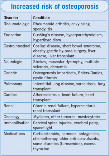 Table 1(click to enlarge)


