CANCER

Obesity, inflammation and cancer

Investigation of anti-inflammatory agents and further understanding of the link between obesity, inflammation and cancer may provide new avenues for the development of anti-cancer treatments

Prof John V Reynolds, Professor of Surgery, Department of Surgery, Trinity Centre for Health Science, St James's Hospital, Dublin 8, Dr Claire L Donohoe, Specialist Registrar in General Surgery, Department of Surgery, Trinity Centre for Health Science, St James’ Hospital, Dublin 8 and Ms Suzanne L Doyle, Dietitian and PhD Research Fellow, Department of Surgery, Trinity Centre for Health Sciences, St James's Hospital, Dublin 8

December 1, 2012

Article
Similar articles
  • Cancers arise as a result of acquired changes in the DNA of cancer cells. Numerous anomalies develop with time and these mutations are positively selected for the micro-environment of the tissue. 

    Obesity is a state of chronic inflammation

    Obesity is characterised by increased storage of fatty acids in an expanded adipose tissue mass.1 Increased adipose tissue mass, especially visceral adipose tissue, is associated with insulin resistance, hyperglycaemia, dyslipidaemia, hypertension and other components of the metabolic syndrome.2 Adipocytes from obese subjects exhibit an altered endocrine function and secretory profile leading to an increased release of pro-inflammatory molecules resulting in a chronic low-grade inflammatory state that has been linked to the development of chronic diseases, including cancer.2

    Despite an expanding body of epidemiological evidence in support of the link between obesity and cancer, the underlying molecular mechanisms responsible are poorly characterised. Excess adipose tissue results in elevated levels of pro-inflammatory adipokines, resulting in an imbalance between increased inflammatory stimuli and decreased anti-inflammatory mechanism leading to persistent low-grade inflammation.3-5

    Adipose tissue is principally deposited in two compartments – subcutaneously and centrally. It is thought that centrally deposited, or visceral, fat is more metabolically active than peripheral subcutaneous fat.1,6,7

    Visceral adipose tissue is largely comprised of omental adipose tissue but also includes other intra-abdominal fat sources, such as mesenteric fat. Visceral adipose tissue secretes a number of adipokines and cytokines leading to a pro-inflammatory, procoagulant and insulin-resistant state, collectively known as the metabolic syndrome.8

    The importance of adipose tissue location in terms of dysmetabolism risk is evident as central obesity is more strongly associated with increased risk of insulin resistance, the metabolic syndrome and cardiovascular diseases than BMI alone.9 For any given amount of total body fat, the subgroup of individuals with excess visceral fat (versus subcutaneous fat) is at higher risk of developing insulin resistance10 and the features of the metabolic syndrome.11

    Visceral fat remains more strongly associated with an adverse metabolic risk profile even after accounting for the contribution of other standard anthropometric indices.12 These systemic effects exerted by visceral adiposity are putatively involved in cancer biology13 and are the focus of current research.

    Inflammation is a cancer hallmark

    Hanahan and Weinberg proposed six hallmarks that define properties that tumours acquire in order to maintain a malignant phenotype, including tissue invasion and metastasis, evasion of apoptosis, sustained angiogenesis, insensitivity to growth inhibitors, limitless replicative potential and self-sufficiency in growth signals.14 Mantovani proposed the addition of a seventh property: an inflammatory micro-environment.15

    Adipose tissue, through the systemic alterations associated with obesity, may support the development of malignant potential in susceptible cells through supporting the other malignant processes within cancer cells, such as invasion and metastases, evasion of apoptosis, promotion of angiogenesis and systemic inflammation. Inflammation provides the selective pressure that may drive the accumulation of mutations and result in poor immune surveillance of tumours once they develop.

    Insulin resistance and obesity

    Obesity is associated with an increased risk of developing insulin resistance and type 2 diabetes. Nutritionally-induced insulin resistance develops as a metabolic adaptation to increased circulating levels of free fatty acids (FFAs), which are constantly released from adipose tissue, especially from visceral fat stores.16

    Increased FFA levels force liver muscle and other tissues to shift towards increased storage and oxidation of fats for their energy production.17 The compensatory effect is a reduced capacity of these tissues to absorb, store and metabolise glucose. Insulin resistance and type 2 diabetes have been recently recognised as intimately associated with the presence of systemic inflammation. There are increased levels of markers and mediators of inflammation including acute phage reactants, such as fibrinogen, C-reactive protein, IL-6, plasminogen activator inhibitor-1 (PAI-1) and white cell count in patients with newly diagnosed type 2 diabetes.18

    Increased insulin in the insulin-resistant state is a mitogen

    Increased numbers of adipocytes releasing FFAs are not alone sufficient for the development of insulin resistance. In addition to increased FFA levels, high concentrations of cytokines produced by adipose tissue, such as TNF-alpha, interleukin (IL) 6 and IL-1beta, and low concentrations of adiponectin are required for deleterious effects on glucose homeostasis.19

    The cellular and molecular mechanisms leading to insulin resistance include a reduction in cellular insulin-receptor levels and reduced responsiveness of some intracellular transduction pathways mediating the effects of insulin binding to its receptor.20 Insulin resistance leads to increased insulin production and insulin can act as a mitogen and has been associated with several cancers.21-23

    The tumorigenic effects of insulin could be directly mediated by insulin receptors in the pre-neoplastic target cells, or might be due to related changes in endogenous hormone metabolism, secondary to hyperinsulinaemia 16

    Adipose tissue in obese individuals is infiltrated by immune cells

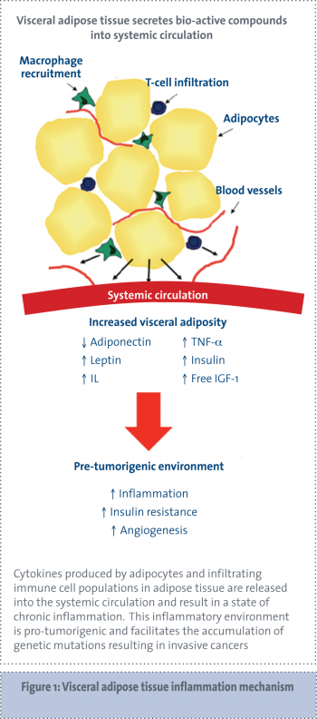
    Cytokines secreted by adipose tissue include the following pro-inflammatory cytokines: TNF-alpha, IL-6, IL-8, IL-10, IL-1 receptor agonist (IL-1Ra), macrophage inflammatory protein 1 (MIP-1) and monocyte chemo-attractant protein 1 (MCP-1). The increased size and number of adipocytes in adipose tissue results in areas of hypoxia within the tissue. This hypoxia induces the secretion of inflammatory factors in order to promote angiogenesis.24-26

    Inflammatory cytokines attract infiltrating immune cells, which in turn produce more inflammatory cytokines. In fact, the overall level of adipokine production from adipose tissue is strongly influenced by the degree of immune cell infiltration present in adipose tissue.6,27-29

    Adipose tissue in obese people is infiltrated with macrophages and the number of macrophages correlates with the degree of adiposity (see Figure 1).30

     (click to enlarge)

    Peripheral monocytes are recruited by MCP-1 and TNF-alpha, and can differentiate into activated macrophages.31 The products of activated macrophages can impact on adipocyte function and are postulated to be involved in altering adipose tissue glucose handling and thus contribute to insulin resistance.32,33

    Research has shown that co-culture of adipocytes with macrophage-conditioned media causes increased adipokine and inflammatory cytokine production by adipocytes,34 further supporting this hypothesis. Increased release of inflammatory cytokines by obese adipose tissue had a profound influence on immune cells both locally and systemically.

    Tumours are ‘wounds that never heal’

    The micro-environment of tumours resembles that of healing wounds. Breaks in epithelium stimulate local acute inflammation, which results in the recruitment of inflammatory cells, fibrin formation and angiogenesis, in order to promote wound healing. If this acute inflammatory process continues, chronic inflammation develops and leads to chronic ulceration or a wound that does not heal. Indeed, French surgeon Jean-Nicolas Marjolin long ago recognised that malignancy can arise in chronic ulcers – indicating that chronic inflammation is carcinogenic.

    The stroma surrounding malignant cells has a similar composition to that of chronically inflamed wounds. In obesity-associated systemic inflammation, there are increased circulating factors that activate the cells of the tumour micro-environment. 

    The cells in the tumour micro-environment (including fibroblasts, neutrophils, T-cells, macrophages and mast cells) secrete factors that support proliferation and invasion of epithelium cells, which under normal circumstances would result in wound healing but, in the presence of mutated cancer cells, promotes their further proliferation and invasion.

    Immune cells can aid cancer cell survival

    While the immune system plays a fundamental role in detecting and eradicating tumour cells under certain circumstances, such as is hypothesised in the obese state, immune cells can aid tumour development and progression.

    The most abundant subsets of infiltrating immune cells within tumours are lymphocytes and macrophages. The density of tumour-associated macrophages (TAMs) is correlated in most studies with increased angiogenesis, tumour invasion and poor prognosis.35

    On one hand, TAMs produce a large number of tumour-promoting factors, including growth factors, matrix metalloproteases and angiogenic factors, such as VEGF. On the other hand, they also produce factors that can suppress adaptive immunity.36 That is, they attract T-cells that are incapable of activating anti-tumour immunity. It is thought that TAMs are derived from circulating monocytes, which are attracted to the tumour site by local production of chemokines.37

    TAMs often accumulate in necrotic areas of tumours, which are characterised by low oxygen tension, and it is thought that hypoxia-induced factors, such as HIF-1α, VEGF, CXCL12 and CXCR4, result in TAM differentiation at the tumour site.38-40

    Obesity is also associated with an alteration in the function of circulating immune cells. Studies have found decreased T-cell and B-cell function, increased monocyte and granulocyte phagocytosis and oxidative burst, and raised total leukocyte counts.41,42 Circulating mononuclear cells from obese subjects have been shown to exhibit increased nuclear factor B (NF-B) nuclear binding with decreased levels of nuclear factor kappa B (NFκB) inhibitor and increased mRNA expression of IL-6, TNF-alpha and migration inhibition factor. Markers of macrophage activation correlate with plasma levels of FFAs.43

    Conclusion

    Thus, altered adipokine production by adipose tissue, and in particular inflamed visceral adipose tissue, may influence the tumour micro-environment in a number of potential ways. 

    Firstly, systemic inflammatory cytokines are increased in obesity. This may affect the tumour micro-environment either directly by stimulating the development of pro-tumorigenic tumour-associated macrophages within the tumour or indirectly, by activating inflammatory pathways in tumour cells which, in turn, stimulate pro-tumorigenic stromal immune cells. 

    Secondly, adipose tissue itself may be the source of pro-tumour immune cells, which migrate to the tumour from inflamed adipose tissue. Finally, systemic inflammation in obesity leads to circulating immune T-cells with decreased anti-tumour activity.

    Animal models provide some evidence supporting these mechanisms. Obese rat models have increased inflammatory transcription factor expression (TNF-alpha and NFκB) in their tumour cells, indicating activation of the tumour cells themselves to produce inflammatory cytokines, which suppress the surrounding immune cells.44

    Furthermore, there is emerging evidence that adipose tissue stromal cells may be a source of stromal cells in tumour micro-environments. Early adipocyte precursor cells can differentiate into stromal cells.45 In animal models of obesity, adipose stromal cells and adipose endothelial cells from inflamed visceral adipose tissue migrate to tumour sites.46 Stromal cells in the tumour micro-environment promote angiogenesis and support tumour progression.47

    Within the clinical setting, there is some evidence that visceral adiposity can influence a patient’s treatment outcome, with a study demonstrating increased visceral fat area to be an independent predictor of outcome after first-line bevacizumab treatment in colorectal cancer.48

    This finding indicates that angiogenic factors produced by visceral fat may influence tumour progression and response to chemotherapy, and is mirrored in animal models as adiponectin, which is reduced in visceral obesity, inhibits tumour growth by reduced neovascularisation.49 The mechanism(s) for this resistance may uncover important information on how obesity influences the tumour micro-environment and further research in this area is warranted. 

    Investigation of anti-inflammatory agents and further understanding of the link between obesity, inflammation and cancer may provide new avenues for the development of anti-cancer treatments.

    Acknowledgements

    This work is funded by an Irish Cancer Society Research Scholarship. The authors declare no conflict of interest.

    References

    1. Galic S, Oakhill JS, Steinberg GR. Adipose tissue as an endocrine organ. Mol Cell Endocrinol 2010; 316: 129-139.
    2. Schroeder DD, Sell H, Uhlig M, et al. Autocrine Action of Adiponectin on Human Fat Cells Prevents the Release of Insulin Resistance-Inducing factors. Diabetes 2005; 54: 2003-2011
    3. Esposito K, Giugliano D. The metabolic syndrome and inflammation: association or causation? Nutr Metab Cardiovasc Dis 2004; 14: 228-232.
    4. Wajchenberg BL. Subcutaneous and visceral adipose tissue: their relation to the metabolic syndrome. Endocr Rev 2000; 21(6): 697-738.
    5. Das UN. Is obesity an inflammatory condition? Nutrition 2001; 17(11-12): 953-966.
    6. Kershaw EE, Flier JS. Adipose tissue as an endocrine organ. J Clin Endocrinol Metab 2004; 89(6): 2548-2556.
    7. Vohl MC, Sladek R, Robitaille J, et al. A survey of genes differentially expressed in subcutaneous and visceral adipose tissue in men. Obes Res 2004; 12(8): 1217-1222.
    8. Despres J-P, Lemieux I. Abdominal obesity and metabolic syndrome. Nature 2006; 444(7121): 881-887.
    9. Nedungadi TP, Clegg DJ. Sexual dimorphism in body fat distribution and risk for cardiovascular diseases. J Cardiovasc Transl Res 2009; 2: 321-327.
    10. Kissebah AH, Vydelingum N, Murray R, et al. Relation of Body Fat Distribution to Metabolic Complications of Obesity. J Clin Endocrinol Metab 1982; 54(2): 254-260.
    11. Despres JP, Moorjani S, Lupien PJ, et al. Regional distribution of body fat, plasma lipoproteins, and cardiovascular disease. Arteriosclerosis 1990; 10(4): 497-511.
    12. Snijder MB, Dam RMV, Visser M, et al. What aspects of body fat are particularly hazardous and how do we measure them? Int J Epidemiol 2006; 35: 83-92.
    13. van Kruijsdijk RCM, van der Wall E, Visseren FLJ. Obesity and Cancer: The Role of Dysfunctional Adipose Tissue. Cancer Epidemiol Biomark Prev 2009; 18(10): 2569-2578.
    14. Hanahan D and Weinberg RA. The hallmarks of cancer. Cell 2000; 100(1): 57-70.
    15. Mantovani A. Cancer: Inflaming metastasis. Nature 2009; 457(7225): 36-37.
    16. Calle EE, Kaaks R. Overweight, obesity and cancer: epidemiological evidence and proposed mechanisms. Nat Rev Cancer 2004; 4: 579–591.
    17. Bergman RN, Ader M. Free fatty acids and pathogenesis of type 2 diabetes mellitus. Trends Endocrinol Metab 2000; 11: 351-356.
    18. Shoelson SE, Lee J, Goldfine AB. Inflammation and insulin resistance. J Clin Invest 2006; 116(7): 1793-1801.
    19. Greenberg AS, McDaniel ML. Identifying the links between obesity, insulin resistance and beta-cell function:potential role of adipocyte-derived cytokines in the pathogenesis of type 2 diabetes. Eur J Clin Invest 2002; 32(Suppl 3): 24–34.
    20. Moller DE, Flier JS. Insulin resistance – mechanisms, syndromes, and implications. NEJM 1991; 325: 938-948.
    21. Trevisan M, Liu J, Muti P, et al. Markers of insulin resistance and colorectal cancer mortality. Cancer Epidemiol Biomark Prev 2001; 10(9): 937-941.
    22. Colangelo LA, Gapstur SM, Gann PH, et al. Colorectal cancer mortality and factors related to the insulin resistance syndrome. Cancer Epidemiol Biomark Prev 2002; 11(4): 385-391.
    23. Schoen RE, Tangen CM, Kuller LH, et al. Increased blood glucose and insulin, body size, and incident colorectal cancer. J Natl Cancer Inst 1999; 91(13): 1147-1154.
    24. Trayhurn P, Wood IS. Adipokines: inflammation and the pleiotropic role of white adipose tissue. Br J Nutr 2004; 92(3): 347-355.
    25. Feldser D, Agani F, Iyer NV, et al. Reciprocal positive regulation of hypoxia-inducible factor 1alpha and insulin-like growth factor 2. Cancer Res 1999; 59(16): 3915-3918.
    26. Fukuda R, Hirota K, Fan F, et al. Insulin-like growth factor 1 induces hypoxia-inducible factor 1-mediated vascular endothelial growth factor expression, which is dependent on MAP kinase and phosphatidylinositol 3-kinase signaling in colon cancer cells. J Biol Chem 2002; 277(41): 38205-38211.
    27. Schaffler A, Muller-Ladner U, Scholmerich J, Buchler C. Role of adipose tissue as an inflammatory organ in human diseases. Endocr Rev 2006; 27(5): 449-467.
    28. Xu H, Barnes GT, Yang Q, et al. Chronic inflammation in fat plays a crucial role in the development of obesity-related insulin resistance. J Clin Invest 2003; 112(12): 1821-1830.
    29. Weisberg SP, McCann D, Desai M, et al. Obesity is associated with macrophage accumulation in adipose tissue. J Clin Invest 2003; 112(12): 1796-1808.
    30. Neels JG, Olefsky JM. Inflamed fat: what starts the fire? J Clin Invest 2006; 116(1): 33-35.
    31. Curat CA, Miranville A, Sengenes C, et al. From blood monocytes to adipose tissue-resident macrophages: induction of diapedesis by human mature adipocytes. Diabetes 2004; 53(5): 1285-1292.
    32. Sartipy P, Loskutoff DJ. Monocyte chemoattractant protein 1 in obesity and insulin resistance. Proc Natl Acad Sci USA 2003; 100(12): 7265-7270.
    33. Wellen KE, Hotamisligil GS. Obesity-induced inflammatory changes in adipose tissue. J Clin Invest 2003; 112(12): 1785-1788.
    34. Bassols J, Ortega FJ, Moreno-Navarrete JM, et al. Study of the proinflammatory role of human differentiated omental adipocytes. J Cell Biochem 2009; 107(6): 1107-1117.
    35. Balkwill F. Cancer and the chemokine network. Nat Rev Cancer 2004; 4(7): 540-550.
    36. Sica A, Allavena P, Mantovani A. Cancer related inflammation: the macrophage connection. Cancer Lett 2008; 267(2): 204-215.
    37. Pollard JW. Tumour-educated macrophages promote tumour progression and metastasis. Nat Rev Cancer 2004; 4(1): 71-78.
    38. Lewis C, Murdoch C. Macrophage responses to hypoxia: implications for tumor progression and anti-cancer therapies. Am J Pathol 2005; 167(3): 627-635.
    39. Talks KL, Turley H, Gatter KC, et al. The expression and distribution of the hypoxia-inducible factors HIF-1alpha and HIF-2alpha in normal human tissues, cancers, and tumor-associated macrophages. Am J Pathol 2000; 157(2): 411-421.
    40. Knowles HJ, Harris AL. Macrophages and the hypoxic tumour microenvironment. Front Biosci 2007; 12: 4298-4314.
    41. Nieman DC, Henson DA, Nehlsen-Cannarella SL, et al. Influence of Obesity on Immune Function. J Am Dietetic Assoc 1999; 99(3): 294-299.
    42. Mendall MA, Patel P, Asante M, et al. Relation of serum cytokine concentrations to cardiovascular risk factors and coronary heart disease. Heart 1997; 78(3): 273-277.
    43. Ghanim H, Aljada A, Hofmeyer D, et al. Circulating Mononuclear Cells in the Obese Are in a Proinflammatory State. Circulation 2004; 110(12): 1564-1571.
    44. Jain SS, Bird RP. Elevated expression of tumor necrosis factor-alpha signaling molecules in colonic tumors of Zucker obese (fa/fa) rats. Int J Cancer 2010; 127(9): 2042-2050.
    45. Rodeheffer MS, Birsoy K, Friedman JM. Identification of White Adipocyte Progenitor Cells In Vivo. Cell 2008; 135(2): 240-249.
    46. Zhang Y, Daquinag A, Traktuev DO, et al. White adipose tissue cells are recruited by experimental tumors and promote cancer progression in mouse models. Cancer Res 2009; 69(12): 5259-5266.
    47. Coussens LM, Werb Z. Inflammation and cancer. Nature 2002; 420(6917): 860-867.
    48. Guiu B, Petit JM, Bonnetain F, et al. Visceral fat area is an independent predictive biomarker of outcome after first-line bevacizumab-based treatment in metastatic colorectal cancer. Gut 2010; 59(3): 341-347.
    49. Brakenhielm E, Veitonmaki N, Cao R, et al. Adiponectin-induced antiangiogenesis and antitumor activity involve caspase-mediated endothelial cell apoptosis. Proc Natl Acad Sci USA 2004; 101(8): 2476-2481.
    © Medmedia Publications/Cancer Professional 2012