The prevalence of atopic dermatitis has increased significantly in industrialised countries and its management presents ongoing challenges
Dr Deirdre O'Callaghan, Dermatology Registrar, Galway University Hospital, Galway and Dr Trevor Markham, Consultant Dermatologist, Galway University Hospital, Galway
Atopic dermatitis (AD or eczema) is a chronic, relapsing, inflammatory skin condition characterised by pruritus and a dry scaly rash. AD may be associated with other atopic diseases such as food allergy, allergic rhinoconjunctivitis and asthma.
The prevalence of AD has increased significantly in industrialised countries in the past three decades, with estimates varying between 10-20% in children and 2-10% in adults.1 It is usually the first manifestation of atopic disease. A total of 40% of cases of AD present within the first six months of life and 85% by the age of five years.2 The disease can also present in adulthood.
Pathophysiology
AD is a multifactorial disease with dysregulation of innate and adaptive immunity based on a strong genetic predisposition. Skin barrier dysfunction contributes to susceptibility to infections and hyper-reactivity to environmental stimuli.3
According to the revised nomenclature of AD, only eczema of patients with elevated total serum IgE (>150kU/l) and specific IgE responses to aero- and food-derived allergens should be considered to have AD.4
Epidermal barrier dysfunction is a prerequisite for the penetration of high-molecular-weight allergens, eg. pollens, microbes, foods etc, inducing inflammation, IgE-mediated sensitisation and potentially leading to the atopic march.5 The initial mechanisms that induce skin inflammation in AD are not known. Is it a primary defect in the innate immune response or a primary defect in the epidermal barrier?
What we know so far
The concordance rate for AD is higher among monozygotic twins (77%) than among dizygotic (15%).6
Genome-wide screens have highlighted several possible susceptibility loci. The region of highest linkage was to the epidermal differentiation complex (EDC) locus on 1q21.7 The EDC is comprised of a cluster of gene families encoding proteins involved in skin cell differentiation.
The filaggrin gene on chromosome 1q21.3 is a key component of the epidermal differentiation complex of the stratum corneum.8 Several loss-of-function mutations of the gene have been identified in AD patients and are associated with patients with high total serum IgE levels and concomitant allergic sensitisations and early age of onset with chronic persistent course until adulthood.9-11
Helper T-cell subsets (TH1, TH2, TH17) have distinct physiological roles in normal skin. In AD there is an imbalance in these T-cell subsets, with TH2 cells predominating in the acute stage resulting in the production of TH2 cytokines 1L-4, 1L-5 and IL-13. In chronic AD, TH1 and TH0 cells (naïve T-cells) predominate, leading to increased interferon-γ, IL-12, IL-5 and GM-CSF.12-13
TH2 cytokines have been shown to downregulate the expression of antimicrobial peptides human cathelicidin LL-37, human β-defensin (HBD)-2 and HBD-3, contributing to the susceptibility of atopic patients to skin infections.14-16
Epidermal dendritic cells have higher surface expression of the high-affinity receptor for IgE (FceRI) compared to non-atopic eczema patients.17
Interleukin-31 (expressed by TH2 cells) and its receptor are overexpressed in the skin of patients with AD. It is thought to be a major factor in the generation of pruritus and has been shown in vitro to be upregulated by exposure to s-staphylococcal exotoxins.18-20 IgE antibodies against self proteins have been identified in a significant number of patients with AD.21-22
Clinical presentation
Figure 1: eczema herpeticum on face(click to enlarge)
Figure 2: eczema herpeticum on neck(click to enlarge)
The clinical manifestations of AD vary with age. In infancy, AD primarily affects the face, scalp and extensor surfaces of the limbs and usually spares the nappy area. During childhood flexural involvement predominates. In adolescence and adulthood, lichenified plaques affect flexures, head and neck.
Dry skin and pruritus are the primary symptoms and these frequently lead to interrupted sleep patterns. AD typically involves periods of disease exacerbation and remission and, although it often improves with age, it can continue into adulthood.
The development of secondary skin infection in AD is common and should be excluded as a possible cause of rapidly worsening eczema, failure to respond to an appropriate topical steroid and emollient regime and may also be associated with systemic symptoms such as fever and malaise.
Bacterial colonisation of the skin by Staphylococcus aureus occurs in more than 90% of patients with AD.5 Group A ß-haemolytic streptococci are also frequently implicated. Such infections may present with typical impetigo or as worsening of existing eczema with erythema, weeping, pustules or honey-coloured crusting.
Viral infections, in particular herpes simplex virus, are more common in patients with AD. This may present as a localised eruption or may be widespread (eczema herpeticum) and associated with constitutional upset. Herpes simplex infection is suggested by clustered vesicles and pustules which are typically umbilicated and/or punched-out erosions which are uniform in appearance and may coalesce to form large denuded areas.
Management
It is vital that patients and parents are educated regarding the disease and its management and that this is reinforced at every consultation. They need to be confident in the daily treatment regime and need to be able to recognise flares and signs of infection. They also need to be able to identify potential trigger factors and adjust their lifestyle accordingly, where practical.
Potential triggers include:
• Irritants and environmental factors – woollen and certain synthetic materials, soap, detergents and perfumed products (including fabric softener) can have an irritant effect on the skin of patients with AD
• Inhalant allergens, eg. house dust mite, pollens, animal dander, moulds, grasses, etc. Should be considered in children with seasonal flares of atopic eczema, children with atopic eczema associated with asthma or allergic rhinitis, and children aged three years or over with atopic eczema on the face, particularly around the eyes23
• Food allergens – cow’s milk, eggs, peanuts, soy, wheat and seafood are some of the most common food allergens in children.
Consider in children less than five years old with moderate-to-severe AD if the child has persistent AD in spite of optimised management and topical therapy and/or the child has a reliable history of an immediate reaction after ingestion of a specific food.23-24
Children with suspected food allergy should be referred for specialist evaluation and investigation, especially if accompanied by systemic features such as vomiting, altered bowel habit or failure to thrive.23 Refer children who follow a cow’s-milk-free diet for longer than eight weeks (for whatever reason) for specialist dietary advice.23
Diets based on unmodified proteins of other species (eg. goat’s milk, sheep’s milk) or partially hydrolysed formulas should not be used in AD for the management of suspected cow’s milk allergy.23
A six-to-eight-week trial of an extensively hydrolysed protein formula or amino acid formula should be offered in place of cow’s milk formula for bottlefed infants under six months of age with moderate-to-severe eczema not controlled with optimal management with emollients and mild, topical steroids. Diets including soya protein can be offered to children aged six months and over with specialist advice.23
Treatment options
A stepped approach should be used, tailoring treatment to severity of the eczema (see Table 3).
Emollients
The mainstay of treatment. Emollients should always be used, even when the skin is clear. Offer a choice of unperfumed emollients to use every day for moisturising, washing and bathing. Aqueous cream should be recommended as a soap substitute only, not as a moisturiser. Best applied when skin is moist, ie. after a five-minute luke-warm bath. They should be prescribed in large quantities, eg. 250-500g/week. Intensive use of emollients will reduce the need for topical steroids.
Topical steroids
Generally, low-potency topical steroids are recommended for use on areas of thinnest skin, such as the face, neck, groin and axillae, except for short-term use for severe flares, eg. three to five days moderate potency for face/neck or seven to 14 days moderate-to-potent preparations for groin/ axillae. Topical steroids should be prescribed for application once or twice daily. Only apply to areas of active eczema. Apply prior to application of emollient. Ointments are generally more potent than cream preparations. Potent topical steroids should not be used in children less than 12 months of age without specialist dermatological advice.23
Topical calcineurin inhibitors
These agents are not first-line therapy and are not recommended for the treatment of mild AD. It is recommended that these agents only be initiated by physicians with a special interest in dermatology. If eczema is not controlled by topical corticosteroids and where there is a serious risk of important adverse effects from further topical corticosteroid use, particularly irreversible skin atrophy, consider23 topical tacrolimus for moderate-to-severe AD in children aged two years and older.
The frequency of application is once to twice daily initially and should then be weaned to twice-weekly maintenance.25,26 Treatment should only be applied to areas of active eczema and never under occlusion without specialist advice.
The most common local side-effect is skin burning and irritation, but this can be minimised by applying the medication only when the skin is dry. Patients should be advised regarding appropriate sun protection, including use of sunscreen.
Systemic corticosteroids
Short courses may be indicated under specialist supervision to gain control in severe disease.
Antihistamines
If sleep disturbance is a major factor a short trial of an age-appropriate sedating antihistamine can be offered to children aged six months and over.23
Wet wrap and medicated dressings23
Occlusive medicated dressings or dry bandages should not be used in the presence of infected eczema. Whole-body occlusive dressings (either wet-wrap therapy or dry bandages or specialised suits) for use with emollients +/- topical corticosteroids is not first-line therapy and should be initiated only by a healthcare professional with specialist dermatology expertise.
Treatment of infection
Patients and parents need to be educated to identify the signs of infection.
Bacterial infections
• Skin swabs are only indicated if micro-organisms other than S. aureus are suspected or if antibiotic resistance is a concern. If recurrent infected eczema is an issue consider a nasal swab to assess nasal carriage of both patients and parents as it is thought parental S. aureus carriage influences S. aureus colonisation of the child.27,28 Topical intranasal mupirocin twice daily for five days of a month intermittently should be considered.29,30
• Localised areas of infection can be treated with a combination topical antibiotic, eg. fusidic acid, and a low-to-medium-potency corticosteroid as appropriate. They should not be used for longer than two weeks.23
• Systemic antibiotics active against S. aureus and streptococcus should be used to treat widespread infection for one to two weeks according to clinical response. Flucloxacillin should be prescribed first line and erythromycin (or clarithromycin) in penicillin-allergic patients.23
• Bleach baths using diluted 2% sodium hypochlorite, eg. Milton, have been shown both anecdotally and in one study (looked at in combination with intranasal mupirocin) to be well tolerated and to decrease the clinical severity of AD in patients with clinical signs of secondary bacterial infections.29 (For instructions on use and dilution see www.eczemacenter.org.)
Herpes simplex infection (HSV)
• If localised HSV skin infection is suspected, treatment with oral aciclovir should be instigated.23
• If widespread HSV infection is suspected (eczema herpeticum), systemic aciclovir should be commenced immediately with same-day referral for specialist dermatology advice.23
• Parents and other close contacts need to be vigilant in treatment of ‘cold sores’ to avoid transmission to patients with eczema.
Phototherapy
• Phototherapy or ultraviolet (UV) treatment is reserved for severe eczema as it is expensive, time-consuming and has potential adverse effects. It involves controlled exposure to UV-B and/or UV-A or PUVA (psoralen plus UVA ), two to three times each week. A treatment course may continue for several months.
• Phototherapy is unsuitable for those who have little flexibility with work or school hours, mobility or transport problems, very fair skin or a history of photosensitivity. It is also unsuitable for very young children as they are unable to stand still for the required period of time and cannot be relied upon to wear safety glasses.
Systemic agents
In patients with severe disease, and particularly in adults, methotrexate, azathioprine, cyclosporin and mycophenolate mofetil have been used with success.
Unproven therapies
Insufficient evidence exists to recommend probiotics or leukotriene inhibitors. The effectiveness and safety of complementary therapies such as homeopathy, massage or traditional Chinese medicines have not been adequately assessed in clinical studies. Parents and patients should be questioned regarding use of over-the-counter or complementary therapies.
Referral criteria
Referral for specialist dermatological advice is recommended if:23
• The diagnosis is, or has become, uncertain
• Moderate-to-severe eczema failing to respond to appropriate therapy
• Suspected contact dermatitis (persistent AD or facial, eyelid or hand atopic eczema)
• Eczema associated with severe or recurrent infections
• Significant psychosocial issues as a consequence of the disease.
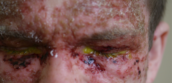 Figure 1: eczema herpeticum on face(click to enlarge)
