CARDIOLOGY AND VASCULAR

Acute AF where pulmonary embolism is driving trigger

There is a common association between atrial fibrillation and pulmonary embolism in clinical practice but not in the literature, write Dr Abuelmagd Abdalla and Dr Tom Walsh

Dr Abuelmagd Abdalla, SHO in Medicine, Wexford General Hospital, Wexford and Dr Tom Walsh, Consultant Geriatrician, University Hospital, Galway

November 2, 2016

Article
Similar articles
  • The case of a 94-year-old woman who had an acute pulmonary embolism (PE) manifested as acute atrial fibrillation (AF) with raised d-dimer and troponin but without other clinical symptoms is presented here. She developed transient AF with rapid ventricular response of 160 beats per minute (bpm), however remained haemodynamically stable without requiring defibrillation or thrombolysis. 

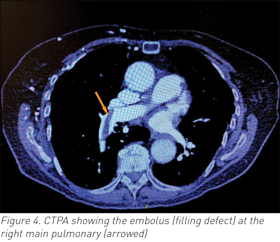
    The underlying PE wasn’t diagnosed when she developed AF. She received timely anticoagulation based on already diagnosed subacute deep vein thrombosis (DVT) and her CT pulmonary angiography (CTPA) subsequently confirmed acute embolus in the right main pulmonary artery. Her AF responded well to beta-blocking therapy. The source was right lower limb DVT. Without AF, her PE would have passed unnoticed. The treatment course would have changed acutely if patient had become unstable, but fortunately this wasn’t the case. This case highlights the possible dilemma of treating acute rapid AF that wasn’t primary but rather secondary to an underlying PE.

    Background

    Pulmonary embolism (PE) is a commonly encountered acute medical emergency. While junior doctors may feel competent to suspect PE in the right clinical setting, it’s becoming more and more complicated and challenging to senior clinicians. There is a spectrum of ECG changes accompanying acute PE, the majority of which are non-specific, especially sinus tachycardia. It’s well known from previous reports that acute right ventricular strain pattern is commonly associated with acute PE.1,2,3 It has also been reported that even ST segment elevation could represent atypical finding in such a setting.4

    Troponin leak has also become a known finding accompanying right ventricular stretching. While the causal relationship between atrial fibrillation (AF) and PE has never been established, it’s a common practice for clinicians in acute medicine to suspect PE with new onset AF in those without significant cardiac history. This case highlights the issue of AF as a proxy to underlying potentially fatal PE.

    Case presentation

    This is a 94-year-old physically well and independent lady who presented with a three week history of right leg swelling that worsened in the past four days, which was confirmed to be due to unprovoked right femoral and popliteal above knee deep vein thrombosis (DVT) by the hospital emergency department. She was admitted medically given her social circumstances and painful leg, and received low molecular weight heparin (LMWH)
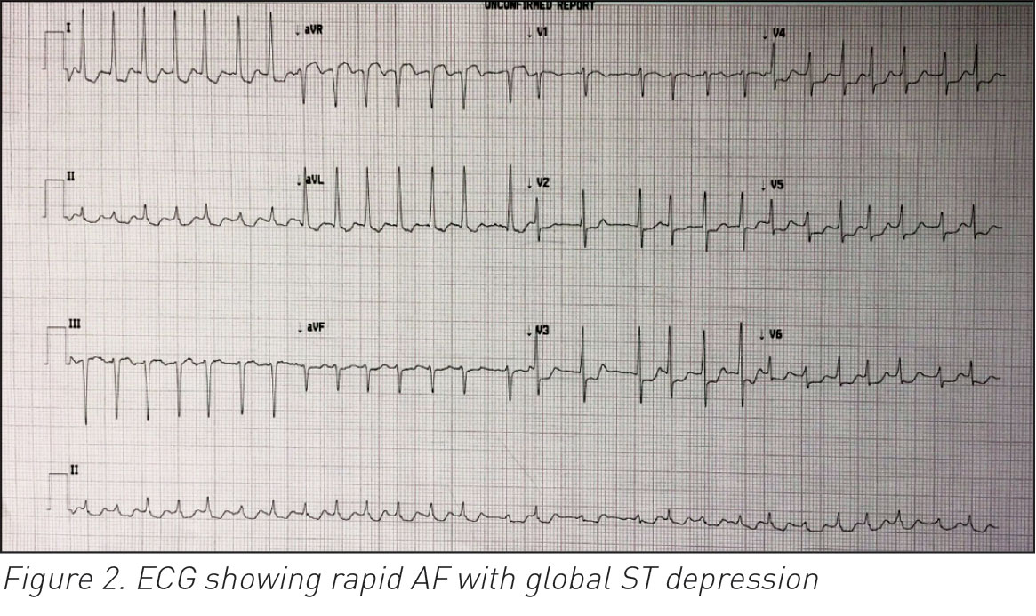
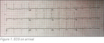
    tinzaparin. She was medically stable and her vitals were within normal on arrival. Her chest was clear of focal signs with no signs of cardiac decompensation but her right thigh and calf were swollen and tender. 

    Her electrocardiogram (ECG) on admission was of sinus rhythm and free of acute ischaemic changes (see Figure 1). Of note is that she denied any chest pain or shortness of breath during the preceding period or her hospital stay. Her past medical history was significant for stable angina, hypertension, osteoporosis and peptic ulcer disease. The following day she suddenly developed acute AF with rapid ventricular response of 150-160 beats per minute (bpm) (see Figure 2)

     (click to enlarge)

     (click to enlarge)

    Her blood pressure (BP) was 115/70, her oxygen saturation was 96% room air and her respiratory rate was 18 breaths per minute. She adequately responded to 2.5mg of intravenous metoprolol and her heart rate settled to 92bpm. Her d-dimer and troponin T were both elevated. She went on to have CT pulmonary angiography (CTPA) 48 hours later which confirmed acute embolus within the right main pulmonary artery. She made slow but good progress over her two weeks of hospitalisation. She was commenced on warfarin and was to remain on same indefinitely. 

    Investigations

    Her bloods on arrival showed elevated d-dimer of over 4,000ng/ml, elevated troponin T of 55ng/L, raised C-reactive protein (CRP) of 19.1mg/L and reduced creatinine clearance at 54ml/minute. Her full blood count indices were normal as well as her coagulation parameters, fibrinogen, creatine kinase (CK), liver function tests (LFTs), urea and electrolytes. Her CRP normalised later. Her chest x-ray was normal. Her ECGs on presentation, during AF and subsequently pre-discharge are illustrated in Figures 1, 2 and 3 respectively. Her CTPA (see Figure 4) showed right central main pulmonary arterial acute embolus. 

     (click to enlarge)

     (click to enlarge)

    Treatment

    She received LMWH tinzaparin on arrival and subsequently warfarin, in addition to bisoprolol and analgesia. She also received input from the multidisciplinary team.

    Discussion

    What is unique about this case is that her PE was totally asymptomatic apart from the episode of transient AF. This could have become detrimental if she was haemodynamically unstable. It would indeed have been challenging for the acute team to decide between defibrillation and thrombolysis if her BP was low or continuing to deteriorate. 

    Bedside echo could also be helpful but its practicalities are not always possible, especially out-of-hours in less staffed hospitals given the timely required medical decision. From a cardiac perspective, with her prior ischaemic heart disease and hypertension history, acute defibrillation for AF is warranted if she is haemodynamically unstable. On the other hand given her DVT, massive PE leading to unstable haemodynamics is a valid possibility. 

    It is interesting to see the agreement rate among the acute team about thrombolysing this patient who otherwise has no major contraindications apart from her age (relative contraindication). AF clinically commonly alerts acute physicians to lower their threshold for suspecting PE diagnosis, however the formal association or any causal relationship has never been established on epidemiological studies. It is well known theoretically that PE increases the right atrial pressure causing stretching injury or dysfunction that can trigger PE. Gex et al in their retrospective cohort of 2,500 found that presence of AF doesn’t increase the probability of PE in patients suspected with this diagnosis5 and this could explain why AF hasn’t yet been part of any PE predicting tool. 

    Flegel6 presented his argument on this matter without arriving at a final conclusion given the lack of evidence and the lack of interest among clinicians for the fact that management with anticoagulation is the key either way. AF as a cause of PE through direct embolisation from right atrial thrombi has also been reported.7 In addition, AF irrespective of its timing, was found to be a prognostic indicator of higher short and medium term mortality following acute PE.8

    Learning points

    • There is a common association between AF and PE in clinical practice but not in the literature
    • Acute clinicians should be extra cautious dealing with acute AF if PE is suspected to be the driving trigger
    • Acute detrimental contradicting decisions may have to be made during clinical instability (eg. defibrillation versus thrombolysis).
    References 
    1. Abarca E, Baddi A, Manrique R. ECG manifestations in submassive and massive pulmonary embolism. Report of 4 cases and review of literature. J Electrocardiol 2014 Jan; [Cited 2015 Dec 25]; 47(1): 75-79. Available from: www.sciencedirect.com/science/article/pii/S0022073613003403
    2. Abecasis J, Monge J, Alberca D, Grenho MF, Arroja I, Aleixo AM. Electrocardiographic presentation of massive and submassive pulmonary embolism. Rev Port Cardiol 2008 May; 27(5): 591-610. [Cited 2015 Dec 25]; 27(5): 591-610. Available from: http://www.ncbi.nlm.nih.gov/pubmed/18717213
    3. Liao YS, Lai CC, Huang SH, Lin SH. Usefulness of electrocardiographic and radiographic changes in the diagnosis of acute pulmonary embolism. QJM 2013 Aug; [Cited 2015 Dec 20]; 106(8): 767-769. Available from: www.ncbi.nlm.nih.gov/pubmed/23458780
    4. Abdalla A, Kelly F. ‘STEMI-like’ acute pulmonary embolism, an unusual presentation. BMJ Case Rep [Internet]. 2014 Nov 20; [Cited 2014 Nov 20]. Available from: http://casereports.bmj.com/content/2014/bcr-2014-206517.full?keytype=refandijkey=yQDOEFwGaPXNTwK
    5. Gex G, Gerstel E, Righini M, LE Gal G, Aujesky D, Roy PM et al. Is atrial fibrillation associated with pulmonary embolism? J Thromb Haemost [Internet]. 2012 Mar [Cited 2015 Dec 20]; 10(3): 347-351. Available from: www.ncbi.nlm.nih.gov/pubmed/22212132
    6. Flegel KM. When atrial fibrillation occurs with pulmonary embolism, is it the chicken or the egg? CMAJ [Internet]. 1999 Apr 20 [Cited 2015 Dec 20]; 160(8): 1,181-1,182. Available from: www.pubmedcentral.nih.gov/articlerender.fcgi?artid=1230273andtool=pmcentrezandrendertype=abstract
    7. Carmichael AJ, Martin AM. Pulmonary embolism: a complication of right atrial thrombi due to atrial fibrillation. J R Soc Med [Internet]. 1991 May [Cited 2015 Dec 29]; 84(5): 313. Available from: www.pubmedcentral.nih.gov/articlerender.fcgi?artid=1293239andtool=pmcentrezandrendertype=abstract
    8. Barra SNC, Paiva LV, Providência R, Fernandes A, Leitão Marques A. Atrial fibrillation in acute pulmonary embolism: prognostic considerations. Emerg Med J [Internet]. 2014 Apr [Cited 2015 Dec 20]; 31(4): 308-312. Available from: www.ncbi.nlm.nih.gov/pubmed/23349355
    © Medmedia Publications/Hospital Doctor of Ireland 2016