GENERAL MEDICINE

Management strategies for saddle pulmonary embolism

A rare case of SPE where initial anticoagulation therapy showed limited improvement, however systemic thrombolysis therapy resulted in significant recovery for the patient

Dr Alvina Zanib, Registrar Infectious Diseases and General Internal Medicine, University Hospital, Limerick and Dr Syed Shabbar Musavi, Consultant in General Internal Medicine, University Hospital, Limerick

April 1, 2024

Article
Similar articles
  • Saddle pulmonary embolism (SPE) is a rare type of acute pulmonary embolism (PE) that can lead to sudden haemodynamic collapse and death. It is defined radiologically as a thrombus spanning the pulmonary artery trunk and can be mistaken for massive PE, which is defined on the basis of haemodynamic instability. 

    Here, we present a case of SPE in a tertiary care setting, providing insights into its clinical presentation and management. The patient, a woman in her 70s, presented with abrupt-onset dyspnoea, chest tightness and dizziness. Elevated D-dimers prompted a CT pulmonary angiography, revealing a saddle thrombus with bilateral emboli. Although initial anticoagulation therapy showed limited improvement, thrombolysis resulted in significant recovery. The patient was subsequently transitioned to a direct oral anticoagulant (DOAC) and discharged with plans for ongoing monitoring. This case highlights the complexities involved in successful management strategies for SPE, offering valuable insights for clinicians.  

    Acute pulmonary embolism

    Acute pulmonary embolism (PE) poses a potential threat to life and ranks as the third most common cause of cardiovascular death, following ischaemic heart disease and stroke.1,2,3,4 It represents the most severe manifestation of venous thromboembolism (VTE), carrying substantial risks of morbidity and mortality. When left untreated, the mortality rate for acute PE can reach as high as 30%. However, with timely diagnosis and appropriate treatment, the death rate for diagnosed and treated PE is approximately 8%.5

    Saddle pulmonary embolism (PE) is a radiological term denoting a large clot that spans the bifurcation of the pulmonary artery trunk, often extending into bilateral main pulmonary arteries.6 It occurs in 2.6% to 5.4% of patients with PE.7,8 The presence of a large central thrombus in saddle PE may cause concern among clinicians, leading them to categorise it as massive PE and admit patients to the intensive care unit (ICU). 

    However, it is important to note that ‘massive PE’ is a haemodynamic definition, indicating any PE accompanied by shock and haemodynamic collapse.9 Therefore, it is inaccurate to label patients with saddle PE as having massive PE, as there are instances where these patients present with stable haemodynamic values and a relatively benign clinical profile. Saddle PE exhibits diverse characteristics with variable clinical features and outcomes. Limited data are available to guide clinicians in the general management of saddle PE. Some small studies have indicated that saddle PE does not necessarily result in an unfavourable clinical outcome, with mortality rates ranging from 4.5% to 16%.8,10

    Case presentation 

    A woman in her 70s presented to the emergency department reporting shortness of breath. In the previous two days she began experiencing a gradual onset of shortness of breath, even at rest, which exacerbated with minimal exertion. She also reported sensations of chest tightness and dizziness. However, she denied experiencing chest pain, orthopnoea, proximal nocturnal dyspnoea, wheezing, ankle swelling, cough or the production of phlegm. Notably, she had recently been discharged from the hospital after being treated for a lower respiratory tract infection, and her recovery was progressing well, with almost complete resolution of infectious symptoms.

    Her medical history included well-controlled type 2 diabetes and hypertension. She reported no recent surgeries. Regular medications comprised oral hypoglycaemic (metformin), an angiotensin receptor blocker (losartan) and aspirin (75mg OD). She denied any family history of clotting disorders. Additionally, she was a non-smoker and non-drinker, and she maintained complete independence in terms of mobility.

    During examination in the ED she was afebrile, but exhibited a rapid heart rate at 116 beats per minute and a blood pressure of 102/78mmHg. Her respiratory rate was 20 breaths per minute and her oxygen saturation was 92% on room air. Chest auscultation revealed clear air entry in both lungs with no additional sounds. The rest of the examination was unremarkable. 

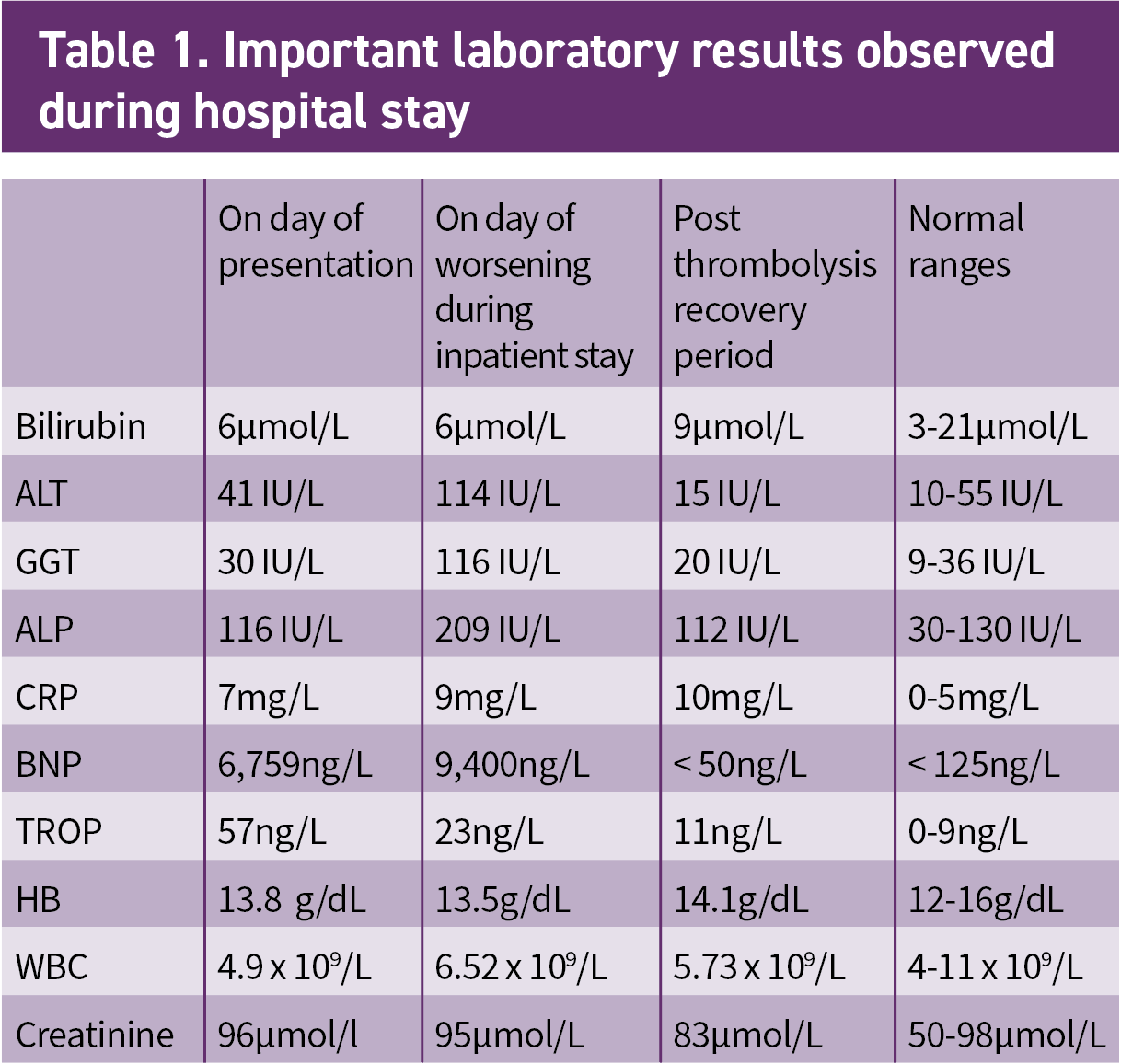
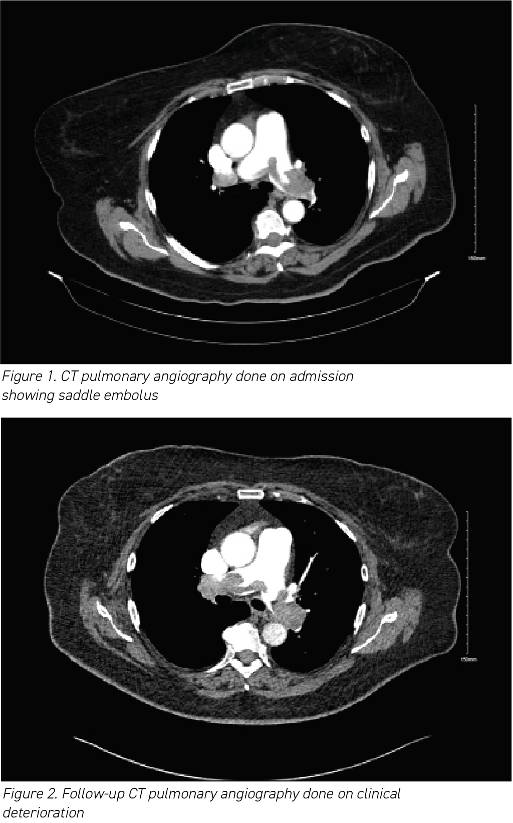
    The results of her routine initial blood investigations are depicted in Table 1. Her initial blood workup revealed elevated D-dimers at > 4.00µg/ml (normal range 0.01 to 1.00µg/ml).

     (click to enlarge)

    Given the elevated D-dimers, a CT pulmonary angiography (CTPA) was conducted, revealing a saddle thrombus in the main pulmonary artery along with massive emboli in both the left and right pulmonary arteries, consistent with a diagnosis of pulmonary thromboembolism (see Figure 1). Subsequently, she was started on a daily regimen of subcutaneous tinzaparin at a dosage of 175 IU/kg.

    During the initial phase of treatment, there was limited improvement in her symptoms and oxygen requirements. Unfortunately, in the subsequent days she experienced further clinical deterioration despite receiving anticoagulation therapy. Her oxygen needs increased to 55-100% Fi02 as shown in Table 2. Although her blood pressure remained stable, it was towards the lower side at 112/53mmHg. To assess the possibility of further clot propagation, additional imaging with a repeat CTPA was performed as the patient was transferred to a high dependency unit.

    The follow-up CTPA (see Figure 2) revealed the persistent presence of a saddle embolus in the main pulmonary artery, along with large-volume bilateral emboli. Interval CT findings indicated evidence of right heart strain, including contrast reflux into the inferior vena cava (IVC), septal straightening and atrial enlargement. Urgent echocardiography was arranged and demonstrated normal left ventricular function, a dilated right ventricle with moderate tricuspid regurgitation, a right ventricular strain pattern and signs of pulmonary hypertension, with a right ventricular systolic pressure of 50mmHg.

     (click to enlarge)

    Given these findings, thrombolysis was considered the most suitable treatment option. She received alteplase with a 10mg bolus followed by a 90mg infusion. Table 2 outlines how her oxygen requirement dropped and blood pressure reading improved post thrombolysis. Concurrently, heparin was administered at a rate of 18 units/kg/hour, with regular monitoring of the activated partial thromboplastin time (APTT). Subsequently, the transition was made to enoxaparin sodium at a dosage of 1mg/kg twice daily, leading to the discontinuation of the heparin infusion.

     (click to enlarge)

    Remarkably, she exhibited significant improvement over the following days. Her oxygen requirements dropped from 55% to 28% and eventually to room air. Additionally, there was an improvement in her blood pressure, which stabilised at 160/80mmHg. Her blood test results demonstrated notable improvement in the subsequent days, as indicated in Table 1.

    Following successful treatment with thrombolysis and initial anticoagulation, she was transitioned to a DOAC, specifically apixaban. Subsequently, she was discharged home with plans for further follow-up at the coagulation clinic to monitor her progress and adjust the treatment regimen as needed.

    Discussion

    In prior research and clinical applications, the term ‘saddle PE’ has frequently been used interchangeably with ‘massive PE’. However, it is important to clarify that ‘massive PE’ is a haemodynamic designation and should exclusively pertain to a pulmonary embolism presenting with significant haemodynamic compromise. In contrast, patients with saddle PE (SPE) can, and frequently do, exhibit entirely stable haemodynamic indicators and minimal symptoms.

    The widespread availability of CT pulmonary angiography (CTPA) has led to an increased recognition of SPE.11,12,13,14 Regarding clinical presentation, dyspnoea and chest pain were the predominant symptoms, consistent with our observed case. Findings from the PIOPED II study indicated that new-onset dyspnoea at rest or during exertion was the most prevalent symptom among patients with pulmonary embolism in general, especially in those without prior cardiopulmonary disease (73%).15 Similarly, the EMPEROR study demonstrated that dyspnoea and chest pain were the most frequently reported initial presentations.16

    The acute occurrence of SPE can induce a sudden elevation in pulmonary arterial pressure and pulmonary vascular resistance, resulting in an increased right ventricular (RV) afterload and subsequent RV dilation, ultimately leading to right-heart failure as was evident in this case. The decrease in RV cardiac output also contributes to a reduction in left ventricular (LV) filling, leading to a decline in blood pressure, which may manifest as syncope, hypotension or cardiogenic shock.5,17 Additionally, acute PE can cause substantial abnormalities in pulmonary gas exchange, leading to hypoxaemia.

    Regarding treatment, it has been observed that patients with SPE receive systemic thrombolytic therapy more frequently than the non-saddle PE group.18 Alkinj B et al found that systemic thrombolysis therapy for late decompensation (occurring > 6 hours after admission) was utilised more often in the saddle PE group compared to the non-saddle PE group (n = 6, 3.2% versus 0%; p = 0.03); similar to this case report.18 These findings also align with the results of Kwak et al19 who identified saddle PE as an independent predictor of major adverse events within 30 days of diagnosis. 

    Clinicians caring for patients with saddle PE should be vigilant about the possibility of delayed decompensation, and it is recommended to closely monitor clinical and haemodynamic parameters within the initial 12 to 24 hours of admission.

    1. Mahar JH, Haddadin I, Sadana D et al. A pulmonary embolism response team (PERT) approach: initial experience from the Cleveland Clinic. J Thromb Thrombolysis 2018 Aug;46(2):186-92
    2. Konstantinides SV, Meyer G, Becattini C et al. Task Force for the diagnosis and management of acute pulmonary embolism of the European Society of Cardiology (ESC) 2019 ESC Guidelines for the diagnosis and management of acute pulmonary embolism developed in collaboration with the European Respiratory Society (ERS). Eur Respir J 2019; Oct 9;54(3):1901647
    3. Raskob GE, Angchaisuksiri P, Blanco AN et al; ISTH Steering Committee for World Thrombosis Day. Thrombosis: a major contributor to global disease burden. Arterioscler Thromb Vasc Biol 2014 Nov;34(11):2363-71
    4. Tagalakis V, Patenaude V, Kahn SR, Suissa S. Incidence of and mortality from venous thromboembolism in a real-world population: the Q-VTE study cohort. Am J Med 2013; 126(9): 832.e13-832.e21
    5. Belohlávek J, Dytrych V, Linhart A. Pulmonary embolism, part I: epidemiology,risk factors and risk stratification, pathophysiology, clinical presentation, diagnosis, and nonthrombotic pulmonary embolism. Exp Clin Cardiol 2013 Spring; 18(2):129-38
    6. Gandara E, Bose G, Erkens P, Rodgers M, Carrier M, Wells P. Outcomes of saddle pulmonary embolism: a nested casecontrol study. J Thromb Haemost 2011; 9(4):867-9
    7. Ryu JH, Pellikka PA, Froehling DA, Peters SG, Aughenbaugh GL. Saddle pulmonary embolism diagnosed by CT angiography: frequency, clinical features and outcome. Respir Med 2007; 101(7):1537-42
    8. Sardi A, Gluskin J, Guttentag A, Kotler MN, Braitman LE, Lippmann M. Saddle pulmonary embolism: is it as bad as it looks? a community hospital experience. Crit Care Med 2011; 39(11):2413-8
    9. Kucher N, Goldhaber SZ. Management of massive pulmonary embolism. Circulation 2005; 112(2):e28-e32
    10. Pruszczyk P, Pacho R, Ciurzynski M et al. Short term clinical outcome of acute saddle pulmonary embolism. Heart 2003; 89(3):335-6
    11. Berdahl CT, Vermeulen MJ, Larson DB, Schull MJ. Emergency department computed tomography utilization in the United States and Canada. Ann Emerg Med 2013; 62(486-94),e3
    12. Stein PD, Fowler SE, Goodman LR et al. Multidetector computed tomography for acute pulmonary embolism. N Engl J Med 2006; Jun 1; 354(22):2317-27
    13. Gottschalk A, Stein PD, Goodman LR, Sostman HD. Overview of prospective investigation of pulmonary embolism diagnosis II. Semin Nucl Med 2002 Jul; 32(3):173-82
    14. Moore AJE, Wachsmann J, Chamarthy MR, Panjikaran L, Tanabe Y, Rajiah P. Imaging of acute pulmonary embolism: an update. Cardiovasc Diagn Ther 2018, Jun; 8(3):225-43
    15. Stein PD, Beemath A, Matta F et al. Clinical characteristics of patients with acute pulmonary embolism: data from PIOPED II. Am J Med 2007; 120(10): 871-9
    16. Pollack CV, Schreiber D, Goldhaber SZ et al. Clinical characteristics, management, and outcomes of patients diagnosed with acute pulmonary embolism in the emergency department: initial report of EMPEROR (Multicenter emergency medicine pulmonary embolism in the real world Registry). J Am Coll Cardiol 2011; 57(6):700-6
    17. Gandara E, Bose G, Erkens P et al. Outcomes of saddle pulmonary embolism: a nested case-control study. J Thromb Haemost 2011 Apr; 9(4):867-9
    18. Alkinj B, Pannu BS, Apala DR, Kotecha A, Kashyap R, Iyer VN. Saddle vs nonsaddle pulmonary embolism: clinical presentation, Hemodynamics, Management, and Outcomes. Mayo Clin Proc 2017; 92(10):1511-8
    19. Kwak MK, Kim WY, Lee CW, Seo DW, Sohn CH, Ahn S, Lim KS, Donnino MW. The impact of saddle embolism on the major adverse event rate of patients with non-high-risk pulmonary embolism. Br J Radiol 2013; 86(1032): 20130273
     
    © Medmedia Publications/Hospital Doctor of Ireland 2024