Modern molecular tools in the treatment of oesophageal cancer
An Irish Cancer Society-funded research fellowship project is focussing on the identification of a unique miRNA expression signature in both tumour tissue and serum predicting therapeutic outcome in oesophageal adenocarcinoma patients.
Dr Stephen G Maher, Senior Research Scientist, Department of Surgery, Institute of Molecular Medicine, Trinity Centre for Health Science, St James's Hospital, Dublin and Prof John V Reynolds, Professor of Surgery, Department of Surgery, Trinity Centre for Health Science, St James's Hospital, Dublin 8
In Ireland, the incidence of oesophageal cancer is rising rapidly. Diagnosis of carcinoma of the oesophagus frequently confers a poor prognosis. In recent times there has been a dramatic epidemiological shift in the histological subtype of oesophageal cancer observed, with adenocarcinoma overtaking squamous cell carcinoma as the predominant cancer form.1 Unfortunately, the overall cure rate is less than 20%, and approximately 40% for localised disease. Consequently, there is great interest in multimodal approaches to therapy and either neoadjuvant chemotherapy alone or combination chemoradiotherapy (CRT) is increasingly the standard of care for locally advanced tumours.2
Within multimodal treatment regimens, the attainment of a complete pathological response to neoadjuvant therapy is a proxy for a favourable outcome, being associated with a five-year survival rate of up to approximately 60%, and more commonly observed with combined CRT compared with chemotherapy alone.3
However, even with CRT such a response is only observed in 20-30% of patients, with the remainder subject to toxicity and increased time to surgery with no apparent gain. It is therefore of significant clinical benefit to identify factors prior to CRT that predict those patients likely to be resistant or sensitive to current regimens. Such an approach may permit more appropriate individualisation of treatment.
The analysis of standard clinicopathological parameters, such as age, sex, obesity status, tumour stage, differentiation and histology, is unable to predict tumour response to CRT.4 There is a wealth of literature evaluating different immunohistochemical protein markers but the data tends to be conflicting and inconclusive. As patients with tumours of similar clinical characteristics can have vastly different responses to CRT, it is possible that this dichotomy is due to subtle differences in the molecular genetic environments of the tumours.
microRNA and cancer
The ability to analyse predictive markers at the level of RNA, DNA and protein has promised to revolutionise our understanding of the disease process, and it is hoped that the era of genomics, transcriptomics and proteomics will herald new biomarkers of response to treatment.
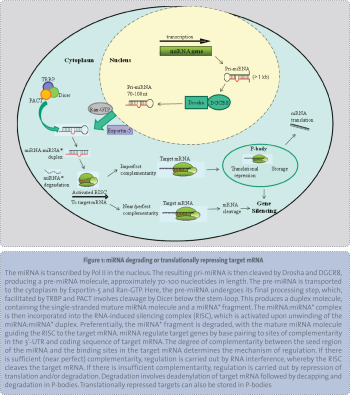
One strategy using gene array technology is to compare the relative gene expression profiles of sensitive and resistant tumours. Coming to the forefront of modern predictive signature research are microRNA (miRNA). Identified in 2001, miRNA are single-stranded RNA molecules that regulate gene expression in cells by directly binding to and either degrading or translationally repressing target mRNA,5(see Figure 1).
(click to enlarge)
Altered miRNA expression is implicated in tumourigenesis and cancer biology.6 A large number of factors may be responsible for the dysregulation of miRNA observed in cancer, including chromosomal aberrations, transcription factor dysregulation, altered or improper processing and epigenetic alterations.
The link between miRNA and cancer was first highlighted when it was discovered that two miRNA, miR-15a and miR-16-1, are located in a region on chromosome 13 that is deleted in over 65% of chronic lymphocytic leukaemia (CLL) patients.7 Despite extensive genetic profiling, no cancer-associated genes had ever been identified within this region, which suggests that miR-15a and miR-16-1 were the genomic targets of this frequent deletion.
Since their original association with cancer, miRNA profiles have been used to discriminate between normal and malignant tissue, in cancers such as lung,8 breast, colorectal,9 pancreatic,10,11 hepatocellular12 and CLL,13 among others. Additionally, it is now possible to delineate and stratify tumours of the same organ of origin, but that have different histologies, for example pulmonary adenocarcinoma and squamous cell carcinoma,8 and endocrine and acinar pancreatic tumours.10
Similarly, miRNA profiles can also be employed as prognostic indicators for factors such as therapeutic outcome and overall survival in cancers such as oesophageal,14 gastric,15 lung,16 osteosarcoma17 and breast.18,19
Genomic tests
Many studies have identified gene expression profiles that are predictive of therapeutic benefit in a variety of cancer types, including breast,20,21 oesophageal22 and colon.23,24 A decade ago, van’t Veer et al20 and van de Vijver et al25 developed the first highly successful predictive gene signature, the so-called Mammaprint, effective in identifying patients with primary breast cancer at a high risk of recurrence after local treatment alone. This signature was independently validated in the MINDACT and TRANSBIG26 trials and was FDA approved in 2007.
Other signatures include the Oncotype DX, a 16-gene signature predicting breast cancer patient response to chemotherapy,27 which was also subsequently validated in a Kaiser Permanente study.28
Conceivably, these stratagems may be applied to oesophageal cancer for predicting response to neoadjuvant CRT, and perhaps in moving forward the future may provide a miRNA-based diagnostic for prognostication in cancer patients.
microRNA as oncogenes/tumour suppressors
While miRNA profiles in cancer are generally tumour-specific, several miRNA are dysregulated across multiple cancers, suggesting a common role in tumourigenesis. miRNA that have been shown to be downregulated in cancers, such as miR-15a, miR-16-1 and the let-7 family, have been proposed to be tumour suppressors, whilst upregulated miRNA such as miR-21 and the miR-17-92 cluster have been classified as oncogenes. miR-21 was one of the first miRNA identified in humans.29 Overexpression of miR-21 has been demonstrated in multiple cancers such as glioblastoma,30 breast,31 oesophageal32 and CLL,33 suggesting a strong oncogenic role for this miRNA. In addition, miR-21-mediated regulation of all three tumour suppressor proteins is associated with increased invasion and metastasis, suggesting a role for miR-21 in cancer progression.
microRNA and cancer therapy
The role of miRNA in the initiation, progression and prognosis of cancer is now well established, however, the role of miRNA in the cellular response to cancer therapy is less well known. Given the role of miRNA in regulating fundamental cellular pathways, such as cell cycle34 apoptosis,35 survival,36 oxidative stress37 and DNA repair,38 it is highly likely that miRNA are involved in the tumour cell responses to anti-cancer therapeutics, which target these key pathways.
In addition to its potential role as an oncogene, miR-21 has also been implicated in the resistance of cancer cells to various chemotherapeutics. miR-21 has been demonstrated to modulate sensitivity to the chemotherapeutic agent doxorubicin in bladder cancer,39 gemcitabine in cholangiocarcinoma40 and 5-Fluorouracil in colorectal cancer.41 The role of miR-21 in both the development and progression of cancer, in addition to the response to anti-cancer treatment, highlights the potential of miR-21 as a novel therapeutic target.
In an isogenic model of resistance to cisplatin in A549 cells, miR-181b was found to be downregulated. Experimental overexpression of miR-181b decreased levels of the anti-apoptotic protein Bcl2, enhancing sensitivity to cisplatin-induced cell death.42
One of the well-established mechanisms involving cisplatin resistance concerns the overexpression of ERCC1. This DNA repair gene is involved in the repair of DNA adducts and stalled DNA replication forks, and its expression levels can predict both survival and cisplatin-based therapeutic benefit in patients with resected non-small cell lung cancer.43,44
Many miRNA appear to be epigenetically silenced by DNA CpG methylation, and as such it may be possible to re-sensitise patients to cisplatin-based chemotherapy through the use of demethylating agents, such as azacitidine or decitabine. Indeed reactivation of genes silenced by methylation of DNA at CpG islands can result in resensitivity to cisplatin in cell line models,45 and such a strategy may work for miRNA.
Radiation has been demonstrated to modulate miRNA expression in cell lines of different tissue origin, including lung cancer,46 lymphoblastoma,47 colon cancer48 and glioma.49 Furthermore, radiation-induced alterations in miRNA expression has been demonstrated to be dose-dependent,37 suggesting that miRNA play a quantifiable, functional role in cellular responses to radiation.
Importantly, a role for miRNA in the in vivo radiation response has been clearly demonstrated. The upregulation of miR-137 and miR-125b in rectal tumour biopsies two weeks after the initiation of neoadjuvant CRT was demonstrated by Svoboda et al, suggesting a role for these miRNA in the tumour response to CRT. Furthermore, increased expression of both miRNA was associated with a poor response to CRT.50
A recent study demonstrated significantly altered expression of just 12 miRNA in resected lung tissue of patients who were resistant and sensitive to adjuvant radiation therapy. One of these miRNA was miR-126, which was upregulated in radiosensitive tumour tissue and was founds to inhibit proliferation and promote radiation-induced apoptosis in vitro.
Potential of microRNA for clinical application
While gene expression profiling has been used in a diagnostic and prognostic capacity, as well as in predicting treatment outcome, generally speaking these approaches have not translated well into a routine clinical setting, for numerous reasons.
For example, most of the techniques require fresh tumour material, or have issues with reproducibility, have complicated bioinformatics due to large data sets, and/or are not cost effective. Circumventing these problems, employing miRNA in diagnostics may be of more efficient clinical utility.
Many investigators now agree that given the direct involvement of miRNA in the regulation of protein expression, miRNA expression profiles may be superior to gene expression profiles for clinical applications, since only a small number of mRNA are regulatory molecules.51
Mirna Therapeutics Inc, a US-based biopharmaceutical biotechnology research and development company, has developed a new type of anti-cancer miRNA technology, which involves using chemically-modified synthetic miRNA mimics and a liposomal-based delivery system to reintroduce a downregulated miRNA back into tumours in vivo.52,53 This strategy may be used to reintroduce miRNA that are important for tumour cell responsiveness to other anti-cancer therapeutics.
It is favourable to a gene therapy approach, as rather than altering a single gene, which will have limited cellular impact, altering a single miRNA will have multiple downstream effects on tumour cells in terms of signalling pathways and effector molecules.
Indeed, a similar strategy has been employed in the treatment of the hepatitis C virus (HCV). It is known that miR-122 is a liver specific miRNA essential for HCV replication.54 It has been demonstrated that HCV replication in the liver can be dramatically reduced using oligonucleotide inhibitors of miR-122.55 This anti-miR-122 (miravirsen) HCV treatment strategy is now in phase II clinical trials. A possibility for the future lies in miRNA replacement therapy for cancer.
Recent findings have shown that miRNAs can also be found in circulation and can be purified from serum/plasma.56-58 Because of their small size miRNAs are highly stable molecules in serum, being protected from cellular RNases, and may be ideal candidates for the development of relatively non-invasive screening tests. In line with this premise, Ng et al recently found five miRNAs in colorectal cancer patients that were upregulated in both plasma and tumour compared to normal controls.59
Irish Cancer Society-funded research
The involvement of miRNAs in the prediction of patient response to therapy in oesophageal cancer remains completely unstudied. Through an Irish Cancer Society-funded research fellowship project our Unit is focussing on the identification of a unique miRNA expression signature in both tumour tissue and serum predicting therapeutic outcome in oesophageal adenocarcinoma patients. Furthermore, we aim to elucidate the functional relevance of specific miRNA in defining responses to radiation and chemotherapy in cell line models of radio- and chemo-resistance. Overall, these data may provide a platform to aid clinicians in deciding whether to send these patients directly for surgery with potential follow up with a course of adjuvant CRT, or to treat with neoadjuvant CRT followed by surgery potentially improving patient prognosis and quality of life.
References
Cook MB, Chow WH, Devesa SS. Oesophageal cancer incidence in the United States by race, sex, and histologic type, 1977-2005. Br J Cancer 2009; 101(5): 855-859
Geh JI, Crellin AM, Glynne-Jones R. Preoperative (neoadjuvant) chemoradiotherapy in oesophageal cancer. Br J Surg 2001; 88(3): 338-356
Chirieac LR, Swisher SG, Ajani JA et al. Post-therapy pathologic stage predicts survival in patients with esophageal carcinoma receiving preoperative chemoradiation. Cancer 2005; 103(7): 1347-1355
Wallqvist A, Rabow AA, Shoemaker RH et al. Linking the growth inhibition response from the National Cancer Institute’s anticancer screen to gene expression levels and other molecular target data. Bioinformatics 2003; 19(17): 2212-2224
Ambros V. The functions of animal microRNAs. Nature 2004; 431(7006): 350-355
Calin GA, Dumitru CD, Shimizu M et al. Frequent deletions and down-regulation of micro- RNA genes miR15 and miR16 at 13q14 in chronic lymphocytic leukemia. Proc Natl Acad Sci U S A 2002; 99(24): 15524-15529
Yanaihara N, Caplen N, Bowman E, Seike et al. Unique microRNA molecular profiles in lung cancer diagnosis and prognosis. Cancer Cell 2006; 9(3): 189-198
Bandres E, Cubedo E, Agirre X et al. Identification by Real-time PCR of 13 mature microRNAs differentially expressed in colorectal cancer and non-tumoral tissues. Mol Cancer 2006; 5: 29
Roldo C, Missiaglia E, Hagan JP et al. MicroRNA expression abnormalities in pancreatic endocrine and acinar tumors are associated with distinctive pathologic features and clinical behavior. J Clin Oncol 2006; 24(29): 4677-4684
Lee EJ, Gusev Y, Jiang J, Nuovo et al. Expression profiling identifies microRNA signature in pancreatic cancer. Int J Cancer 2007; 120(5): 1046-1054
Murakami Y, Yasuda T, Saigo K et al. Comprehensive analysis of microRNA expression patterns in hepatocellular carcinoma and non-tumorous tissues. Oncogene 2006; 25(17): 2537-2545
Calin GA, Liu CG, Sevignani C et al. MicroRNA profiling reveals distinct signatures in B cell chronic lymphocytic leukemias. Proc Natl Acad Sci U S A 2004; 101(32): 11755-11760
Feber A, Xi L, Pennathur A et al. MicroRNA prognostic signature for nodal metastases and survival in esophageal adenocarcinoma. Ann Thorac Surg 2011; 91(5): 1523-1530
Liu R, Zhang C, Hu Z et al. A five-microRNA signature identified from genome-wide serum microRNA expression profiling serves as a fingerprint for gastric cancer diagnosis. Eur J Cancer 2011; 47(5): 784-791
Bianchi F, Nicassio F, Marzi M et al. A serum circulating miRNA diagnostic test to identify asymptomatic high-risk individuals with early stage lung cancer. EMBO Mol Med 2011; 3(8): 495-503
Gougelet A, Pissaloux D, Besse A et al. Micro-RNA profiles in osteosarcoma as a predictive tool for ifosfamide response. Int J Cancer 2011; 129(3): 680-690
Janssen EA, Slewa A, Gudlaugsson E et al. Biologic profiling of lymph node negative breast cancers by means of microRNA expression. Mod Pathol 2010; 23(12): 1567-1576
Iorio MV, Casalini P, Tagliabue E et al. MicroRNA profiling as a tool to understand prognosis, therapy response and resistance in breast cancer. Eur J Cancer 2008; 44(18): 2753-2759
van’t Veer LJ, Dai H, van de Vijver MJ et al. Gene expression profiling predicts clinical outcome of breast cancer. Nature 2002; 415(6871): 530-536
Knauer M, Mook S, Rutgers EJ et al. The predictive value of the 70-gene signature for adjuvant chemotherapy in early breast cancer. Breast Cancer Res Treat 2010; 120(3): 655-661
Maher SG, Gillham CM, Duggan SP et al. Gene expression analysis of diagnostic biopsies predicts pathological response to neoadjuvant chemoradiotherapy of esophageal cancer. Ann Surg 2009; 250(5): 729-737
Del Rio M, Molina F, Bascoul-Mollevi C et al. Gene expression signature in advanced colorectal cancer patients select drugs and response for the use of leucovorin, fluorouracil, and irinotecan. J Clin Oncol 2007;25(7): 773-780
Yi JM, Dhir M, Van Neste L et al. Genomic and epigenomic integration identifies a prognostic signature in colon cancer. Clin Cancer Res 2011; 17(6): 1535-1545
van de Vijver MJ, He YD, van’t Veer LJ et al. A gene-expression signature as a predictor of survival in breast cancer. N Engl J Med 2002; 347(25): 1999-2009
Buyse M, Loi S, van’t Veer L et al. Validation and clinical utility of a 70-gene prognostic signature for women with node-negative breast cancer. J Natl Cancer Inst 2006; 98(17): 1183-1192
Cobleigh MA, Tabesh B, Bitterman P et al. Tumor gene expression and prognosis in breast cancer patients with 10 or more positive lymph nodes. Clin Cancer Res 2005; 11(24 Pt 1): 8623-8631
Habel LA, Shak S, Jacobs MK et al. A population-based study of tumor gene expression and risk of breast cancer death among lymph node-negative patients. Breast Cancer Res 2006; 8(3): R25.
Cai X, Hagedorn CH, Cullen BR. Human microRNAs are processed from capped, polyadenylated transcripts that can also function as mRNAs. RNA 2004; 10(12): 1957-1966
Chan JA, Krichevsky AM, Kosik KS. MicroRNA-21 is an antiapoptotic factor in human glioblastoma cells. Cancer Res 2005; 65(14): 6029-6033
Iorio MV, Ferracin M, Liu CG et al. MicroRNA gene expression deregulation in human breast cancer. Cancer Res 2005; 65(16): 7065-7070
Feber A, Xi L, Luketich JD et al. MicroRNA expression profiles of esophageal cancer. J Thorac Cardiovasc Surg 2008; 135(2): 255-260, discussion 260
Fulci V, Chiaretti S, Goldoni M et al. Quantitative technologies establish a novel microRNA profile of chronic lymphocytic leukemia. Blood 2007; 109(11): 4944-4951
Linsley PS, Schelter J, Burchard J et al. Transcripts targeted by the microRNA-16 family cooperatively regulate cell cycle progression. Mol Cell Biol 2007; 27(6): 2240-2252
Cimmino A, Calin GA, Fabbri M et al. miR-15 and miR-16 induce apoptosis by targeting BCL2. Proc Natl Acad Sci U S A 2005; 102(39): 13944-13949
Meng F, Henson R, Wehbe-Janek H et al. The MicroRNA let-7a modulates interleukin-6-dependent STAT-3 survival signaling in malignant human cholangiocytes. J Biol Chem 2007; 282(11): 8256-8264
Simone NL, Soule BP, Ly D et al. Ionizing radiation-induced oxidative stress alters miRNA expression. PLoS One 2009; 4(7): e6377
Crosby ME, Kulshreshtha R, Ivan M, Glazer PM. MicroRNA regulation of DNA repair gene expression in hypoxic stress. Cancer Res 2009; 69(3): 1221-1229
Tao J, Lu Q, Wu D et al. microRNA-21 modulates cell proliferation and sensitivity to doxorubicin in bladder cancer cells. Oncol Rep 2011; 25(6): 1721-1729
Meng F, Henson R, Lang M et al. Involvement of human micro-RNA in growth and response to chemotherapy in human cholangiocarcinoma cell lines. Gastroenterol 2006; 130(7): 2113-2129
Valeri N, Gasparini P, Braconi C et al. MicroRNA-21 induces resistance to 5-fluorouracil by down-regulating human DNA MutS homolog 2 (hMSH2). Proc Natl Acad Sci U S A 2010; 107(49): 21098-21103
Zhu W, Shan X, Wang T et al. miR-181b modulates multidrug resistance by targeting BCL2 in human cancer cell lines. Int J Cancer 2010; 127(11): 2520-2529
Cobo M, Isla D, Massuti B et al. Customizing cisplatin based on quantitative excision repair cross-complementing 1 mRNA expression: a phase III trial in non-small-cell lung cancer. J Clin Oncol 2007; 25(19): 2747-2754
Allingham-Hawkins D, Lea A, Levine S. ERCC1 Expression Analysis to Guide Therapy in Non-Small Cell Lung Cancer. PLoS Curr 2010; 2: RRN1202
Chang X, Monitto CL, Demokan S et al. Identification of hypermethylated genes associated with cisplatin resistance in human cancers. Cancer Res 2010; 70(7): 2870-2879
Shin S, Cha HJ, Lee EM et al. Alteration of miRNA profiles by ionizing radiation in A549 human non-small cell lung cancer cells. Int J Oncol 2009; 35(1): 81-86
Cha HJ, Shin S, Yoo H et al. Identification of ionizing radiation-responsive microRNAs in the IM9 human B lymphoblastic cell line. Int J Oncol 2009; 34(6): 1661-1668
Shin S, Cha HJ, Lee EM et al. MicroRNAs are significantly influenced by p53 and radiation in HCT116 human colon carcinoma cells. Int J Oncol 2009; 34(6): 1645-1652
Chaudhry MA, Sachdeva H, Omaruddin RA. Radiation-induced micro-RNA modulation in glioblastoma cells differing in DNA-repair pathways. DNA Cell Biol 2010; 29(9): 553-561
Svoboda M, Izakovicova Holla L, Sefr R et al. Micro-RNAs miR125b and miR137 are frequently upregulated in response to capecitabine chemoradiotherapy of rectal cancer. Int J Oncol 2008; 33(3): 541-547
Cowland JB, Hother C, Gronbaek K. MicroRNAs and cancer. APMIS 2007; 115(10): 1090-1106
Trang P, Wiggins JF, Daige CL et al. Systemic delivery of tumor suppressor microRNA mimics using a neutral lipid emulsion inhibits lung tumors in mice. Mol Ther 2011; 19(6): 1116-1122
Wiggins JF, Ruffino L, Kelnar K et al. Development of a lung cancer therapeutic based on the tumor suppressor microRNA-34. Cancer Res 2010; 70(14): 5923-5930
Roberts AP, Lewis AP, Jopling CL. miR-122 activates hepatitis C virus translation by a specialized mechanism requiring particular RNA components. Nucleic Acids Res 2011; 39(17): 7716-7729
Young DD, Connelly CM, Grohmann C, Deiters A. Small molecule modifiers of microRNA miR-122 function for the treatment of hepatitis C virus infection and hepatocellular carcinoma. J Am Chem Soc 2010; 132(23): 7976-7981
Chen X, Ba Y, Ma L et al. Characterization of microRNAs in serum: a novel class of biomarkers for diagnosis of cancer and other diseases. Cell Res 2008; 18(10): 997-1006
Lawrie CH, Gal S, Dunlop HM et al. Detection of elevated levels of tumour-associated microRNAs in serum of patients with diffuse large B-cell lymphoma. Br J Haematol 2008; 141(5): 672-675
Mitchell PS, Parkin RK, Kroh EM et al. Circulating microRNAs as stable blood-based markers for cancer detection. Proc Natl Acad Sci U S A 2008; 105(30): 10513-10518
Ng EK, Chong WW, Jin H et al. Differential expression of microRNAs in plasma of patients with colorectal cancer: a potential marker for colorectal cancer screening. Gut. 2009; 58(10): 1375-1381
 (click to enlarge)