GENERAL MEDICINE

PET – latest techniques in nuclear imaging

PET has fundamentally changed not only the assessment and treatment of patients but how we conceptualise disease itself

Dr Leo P Lawler, Consultant Radiologist, Mater Misericordiae, University Hospital, Dublin

January 1, 2011

Article
Similar articles
  • Though it was born in the 1970s most of us did not see positron emission tomography (PET) in clinical practice until the late 1990s. Given the very complex and expensive technology involved it was initially confined to those units that were heavily involved in its development. 

    My first encounter with PET was in such a unit at the Mallinckrodt Institute of Technology, St Louis, Missouri, in the US. But rather like the iPhone and iPad, it arrived with a combination of high impact and utility such that quickly few of us could understand how we worked without it. 

    The major role of PET in the diagnostic evaluation process was universally acknowledged and its momentum gathered pace so that combined PET-CT (PET-computed tomography) today is very much a mainstream tool in radiology practice. Manufacturers have helped ensure the technology is now accessible to average-sized hospitals and imaging centres. 

    This technology has fundamentally changed not only how we assess patients and treat them but how we conceptualise the disease itself. It has shifted modern imaging development of superlative anatomic detail into the realm of metabolic and functional assessment of the body. In this paper we shall discuss a brief overview of PET-CT practice and then discuss its major applications in oncological and non-oncological diseases processes in 2011.

    What is PET-CT?

    PET is a particular nuclear medicine imaging tool that utilises positron (an electron-like particle but with a positive charge) emitting radionuclide agents. At their site of uptake they emit two photon rays (called gamma rays) 180 degrees opposite to one another. This permits an image reconstruction reflecting the distribution of activity even though the sites of uptake are in very small picomolar quantities. 

    Such PET radionuclides include carbon, nitrogen, oxygen, fluorine, galium and rubidium which can be used to label most biomolecules without changing their constitution or properties. 

    F-fluorodeoxyglucose (FDG) is the only agent used commonly in clinical practice as it is easily produced and its relatively long half-life permits ease of distribution. FDG has been proven to be of high utility and broad application and is now firmly part of medical vernacular among all staff grades. 

    This agent is administered intravenously and taken up and trapped within tissues according to their glucose utilisation. Diseased tissues tend to have a high rate of glucose metabolism compared to background tissues.

    At a cellular level many enzymatic changes have been documented including increased glucose transport rates and phosphorylation and decreased de-phosphorylation. 

    We can get an indirect measure of FDG uptake (standardised uptake value; SUV) which can be compared between scans on individual patients and between patients. This quantification is becoming increasingly important in tissue differentiation and assessing tissue biology and response.

    Modern multi-slice CT

    Though PET machines (see Figure 2) were originally stand-alone, all modern PET platforms combine with computed tomography (CT) scanners (PET-CT). Such hybrid imaging was of course a reinvention of two wheels capitalising on the synergy of each modalities’ respective strengths. Modern multi-slice CT technology provides superb high resolution anatomic detail in three dimensions as well as providing a means to improve the PET image quality (attenuation correction). 

    PET-CT is easily performed as an outpatient study. Quality imaging does require a specialist team of radiologist, radiographer, nursing and support staff. There is a specific patient preparation process and patients may receive conventional iodinated contrast as well as FDG. 

    The protocols between patients are broadly similar but there may be some variation to reflect the disease process under study. Behind the scenes there is a large amount of work involved in regulatory compliance and safety given the use of radiation and radionuclides on patients.

    Ireland is fortunate to already have a number of PET-CT centres with superbly trained individuals providing high quality care with this latest technology equivalent to anywhere in the world.

    Role of PET-CT

    The major role of PET-CT internationally in current radiology practice is in cancer care. It also has an emerging role in neurological, cardiac and inflammatory diseases. In specialised PET-CT centres its role may extend into other limited areas and disease states, but the more common and widely accepted roles are discussed here.

    Oncology

    The tools (eg. radiography, ultrasound; US, CT, magnetic resonance imaging; MRI, bone scan) available in most imaging departments in Ireland now suffice to provide a very comprehensive, detailed and accurate assessment of most cancers presenting in hospital practice. 

    Indeed not all cancers need PET-CT but rather PET-CT is applied when one thinks its unique means of characterising a disease state will effect a management change through tissue sampling, treatment or prognosis. 

    Fortunately PET-CT seems to work best in those cancers commonly seen in our society including: lung (see Figure 4), breast, lymphoma (see Figure 3), gastrointestinal (GI; colorectal, oesophageal; see Figure 1), head and neck, gynaecological (uterus, cervix, ovary), melanoma as well as some less common tumours. 

    Though there are broad agreements there are as yet no universally accepted algorithms for the use of PET-CT. For guidance, practitioners look to evidence-based studies, consensus statements from professional bodies, specialty journal recommendations and US insurer/medicare policy. The who, when and why questions are always difficult when such population guidance is filtered down to individual patients. 

    It has a role in lesion characterisation, diagnosis, tissue sampling, staging and assessment of treatment response. 

    PET-CT is not commonly used to diagnose cancer, in fact the majority of patients that present for scanning already have a suggested diagnosis. In such cases it mainly serves to further characterise the primary disease through its activity, location and pattern etc. 

    In select conditions PET-CT can help differentiate benign and malignant lesions eg. a solitary lung nodule and non-small cell lung cancer (NSCLC). PET-CT has been used to search for primary lesions in patients traditionally diagnosed as ‘adenocarcinoma of unknown primary’. 

    In exceptional circumstances PET-CT has been employed to search for a primary oncological process in a patient with a highly suggestive clinical syndrome where traditional methods have failed to yield a causative lesion. Finally, as with the advent of all new imaging technologies PET-CT has brought with it, its own so-called ‘incidentalomas’. This is where the PET-CT is done to evaluate a specific known cancer and yet incidentally picks up a separate synchronous primary lesion. 

    In some lesions when tissue diagnosis is required the PET-CT can be of value. The areas of greatest FDG avidity can be used to guide the operator to the site most likely to yield active tumour cells decreasing sampling error. Examples would include sampling of large tumours with extensive areas of necrosis where the lesion is outgrowing its blood supply. Another is in large infiltrating lung lesion centred in collapsed lung where the activity can help delineate the site of lesion versus that of adjacent collapsed lung. 

    Finally, depending on the individual case, local practice and management algorithms multidisciplinary teams will on occasion accept PET-CT features as pathognomonic for primary and metastatic disease and obviate the need for tissue sampling altogether. 

    For example some centres will operate on an isolated lung lesion which has morphologic and PET features of bronchogenic carcinoma in the absence of tissue given the test’s high positive predictive value.

    Figure 3. Body scan showing lymphoma
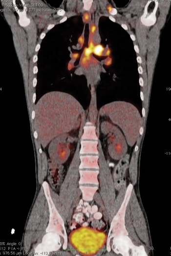
    Figure 3. Body scan showing lymphoma(click to enlarge)

    Figure 2. Patient undergoing PET scan
    Figure 2. Patient undergoing PET scan(click to enlarge)

    Figure 1. Oesophageal carcinoma.
    Figure 1. Oesophageal carcinoma.(click to enlarge)

    CT and MRI sensitivity

    Normal-sized lymph nodes may be cancerous and enlarged lymph nodes may contain no viable tumour cells after therapy. An extranodal soft tissue mass or nodule is not necessarily a cancer. A cancerous mass that doesn’t change with treatment may yet have responded. 

    Some cancerous disease is below the sensitivity of the latest CT and MRI. These are examples of case scenarios where combined evaluation of morphology and FDG-avidity may improve specificity in staging. 

    The high contrast of PET may increase the conspicuity of small lesions such as early liver or bone deposits where the lesion has not yet caused enough end-organ change to declare itself by morphology alone. The pattern of FDG distribution may suggest an alternate diagnosis eg. mediastinal node enlargement in a cancer patient with sarcoid. The PET-CT pattern of lymph nodes may suggest a benign process. Equivocal pleural effusions on CT may be malignant if hot on PET. 

    If a patient already has an advanced stage of disease by standard CT, MRI etc. the PET-CT is unlikely to add to the specifics of the disease state, but may provide a baseline to assess response to therapy. In particular, when there is a mixed response this is quite useful.

    If a patient is surgically unresectable by conventional imaging PET-CT will add little, however, it can prevent futile laparotomies in those deemed resectable by finding metastatic disease occult standard US/CT/MRI. 

    ‘Downstaging’ advanced cancers to the point of being resectable is a controversial area. It assumes that PET-CT sees everything, which of course every cell biologist will tell you is foolish. 

    The premise is that a patient who presents with unresectable disease by PET-CT criteria and shows a complete response to first line chemotherapy by subsequent PET-CT should then be ‘restaged’ and considered for resection of the primary. The jury is still largely out on this fraught issue, but it is clearly entering the debate in practice.1-5

    Figure 4. Scan of patient’s lung
    Figure 4. Scan of patient’s lung(click to enlarge)

    Cardiovascular

    Cardiovascular diagnosis and therapy has traditionally been based on angiographic imaging of macrovascular lumen changes ie. stenosis and occlusion. But PET-CT and other modalities have instead sought to image the vessel wall itself as well as end-organ microvascular change and altered tissue perfusion. In the last decade, attention has been directed to imaging arterial plaque itself during its ongoing dynamic changes, eg. carotid plaque in transient ischaemic attack (TIA), to assess for sites of plaque rupture and instability. 

    With chronic medium vessel vasculitis such as Takayasu disease the SUV of uptake in vessel walls may provide a marker of disease activity and response to therapy.

    Conventional cardiac nuclear medicine and MRI have provided very useful tools for myocardial imaging, but remain under utilised. PET-CT can provide information on stunned, hibernating and infarcted myocardium to assess the role for revascularisation of viable myocardium. 

    FDG is still the commonest cardiovascular imaging tracer, but there are some very short half-life tracers than can be applied with some, but the necessity of local cyclotron production has precluded their use in routine practice.6-10 

    Neuro-imaging

    Functional MRI perhaps is best known for its attempts to unlock brain disease patterns and diagnoses, but there is a steady growth in brain PET-CT for neuropsychiatric disorders in clinical practice. Novel areas include dementia and epilepsy imaging. Again FDG dominates most practices, but there are 15O labels of water, CO2 and oxygen that have been applied to perfusion and oxygen extraction studies. 

    The pattern of distribution of hypometabolism can help differentiate among various dementias, for example:

    • Degenerative eg. Alzheimer’s, Parkinson’s, Huntington’s
    • Vascular eg. multiple infarct
    • Trauma
    • Infection eg. HIV. 

    PET-CT has shown potential in identifying Alzheimer’s disease even before clinical manifestations and structural change. The hope is that early identification will not simply be a lead-time bias, but in fact will permit early interventions that will alter the natural history of the disease. Epilepsy is more challenging with less specific findings and more overlap with normal studies. However, it is suggested that in those patients in whom standard structural MRI studies have not revealed an epileptogenic focus that PET-CT may help lateralise such foci or localise hypometabolic foci in temporal and extratemporal seizures. When positive, such studies may predict response to surgical procedures, but they have to be timed and correlated with ictal and postictal phases.

    Inflammatory imaging

    Though not initially designed for inflammation it was clear after a period in use that this was going to be an adjunct PET-CT application. Most commonly abnormal inflammatory conditions are an incidental finding, however, the author and others have applied it to assess disease activity in vasculitis (see previous) and retroperitoneal fibrosis.

    It can help detection, give a measure of disease activity and assess response to therapy. As in traditional indium-based techniques leukocytes may be labelled but this is not as yet superior to FDG alone.9, 11-16

    Basic research

    Both university and industry have invested in animal PET-CT for basic research beyond the clinical realm. For many it provides a new in vivo avenue to understand molecular pathways. 

    Drug development is perhaps one area that has been most explored, in particular assessing target binding by putative new agents to allow researchers accept or reject drugs early in their development pathway. Equally, there is the possibility to develop animal models for disease processes that can be studied without sacrifice. There is clear potential for PET-CT to bridge the clinical research divide with translational endeavour.

    Conclusion

    PET-CT is very much part of routine radiology practice in Ireland and abroad. It is not perfect and we are likely only experiencing its earliest iterations and yet it has had a massive impact on patient care algorithms. I anticipate rapid developments over the next decade in hardware (machines to acquire the pictures), software (to process and interrogate the data acquired) as well as developments in radiotracer agents. 

    Technological refinements and translational research with health technology assessment will ensure that more disease and patient-specific protocols will continue to evolve with greater universality of practice. 

    We can expect to see a slower, but continued growth in non-cancer areas. Already it is effecting change in care pathways and critical decision making at oncology multidisciplinary team meetings. Cost and acquisition time will remain a significant rate limiting steps to access and diffusion of this technology in the short term. 

    There will be parallel developments in other hybrid platforms like PET-MRI and fusion software. PET-CT will help drive other molecular imaging technologies that move us closer to a more complete picture of phenotypic expression of disease looking at both form and function. 

    We shall likely learn more about the futility of some of our therapeutic endeavours as well as where we can better direct our energies for care and cure. As with all imaging tools it will provide best care for patients in centres where radiology is considered part of clinical practice and imaging results are discussed in multidisciplinary team forums with background imaging department audit and quality assessment.

    References

    1. Lee HY, Han J, Lee KS et al. Lung adenocarcinoma as a solitary pulmonary nodule: prognostic determinants of CT, PET, and histopathologic findings. Lung Cancer 2009; 66: 379-85
    2. Pennant M, Takwoingi Y, Pennant L et al. A systematic review of positron emission tomography (PET) and positron emission tomography/computed tomography (PET/CT) for the diagnosis of breast cancer recurrence. Health Technol Assess 2010; 14: 1-103
    3. Langer A. A systematic review of PET and PET/CT in oncology: a way to personalize cancer treatment in a cost-effective manner? BMC Health Serv Res 2010; 10: 283
    4. Muijs CT, Beukema JC, Pruim J et al. A systematic review on the role of FDG-PET/CT in tumour delineation and radiotherapy planning in patients with esophageal cancer. Radiother Oncol 2010; 97: 165-71
    5. Chowdhury FU, Shah N, Scarsbrook AF, Bradley KM. (18F) FDG PET/CT imaging of colorectal cancer: a pictorial review. Postgrad Med J 2010; 86: 174-82
    6. Flotats A, Knuuti J, Gutberlet M et al. Hybrid cardiac imaging: SPECT/CT and PET/CT. Eur J Nucl Med Mol Imaging 2011; 38: 201-12
    7. Saam T, Rominger A, Wolpers S et al. Association of inflammation of the left anterior descending coronary artery with cardiovascular risk factors, plaque burden and pericardial fat volume: a PET/CT study. Eur J Nucl Med Mol Imaging 2010; 37: 1203-12
    8. Knaapen P, de Haan S, Hoekstra OS et al. Cardiac PET-CT: advanced hybrid imaging for the detection of coronary artery disease. Neth Heart J 2010; 18: 90-8
    9. Graebe M, Borgwardt L, Hojgaard L et al. When to image carotid plaque inflammation with FDG PET/CT. Nucl Med Commun 2010; 31: 773-9
    10. Czihal M, Tato F, Forster S et al. Fever of unknown origin as initial manifestation of large vessel giant cell arteritis: diagnosis by colour-coded sonography and 18-FDG-PET. Clin Exp Rheumatol 2010; 28: 549-52
    11. Jasper N, Dabritz J, Frosch M et al. Diagnostic value of [(18)F]-FDG PET/CT in children with fever of unknown origin or unexplained signs of inflammation. Eur J Nucl Med Mol Imaging 2010; 37: 136-45
    12. Ferda J, Ferdova E, Zahlava J et al. Fever of unknown origin: a value of (18)F-FDG-PET/CT with integrated full diagnostic isotropic CT imaging. Eur J Radiol 2010;  73: 518-25
    13. Dong MJ, Zhao K, Liu ZF et al. A meta-analysis of the value of fluorodeoxyglucose-PET/PET-CT in the evaluation of fever of unknown origin. Eur J Radiol 2010
    14. Andres E, Federici L, Imperiale A. Value of 18 FDG-PET/CT in clinical practice in patients with fever of unknown origin and unexplained prolonged inflammatory syndrome. Eur J Radiol 2010; 75: 122
    15. Chavaillaz O, Gueddi S, Taylor S et al. Giant cell arteritis mimicking fever of unknown origin: potential diagnostic role of PET scan. Thromb Haemost 2006; 95:  390-2
    16. Kobayashi Y, Ishii K, Oda K et al. Aortic wall inflammation due to Takayasu arteritis imaged with 18F-FDG PET coregistered with enhanced CT. J Nucl Med 2005; 46: 917-22
    © Medmedia Publications/Modern Medicine of Ireland 2011