CANCER

Sarcoma review: Types, diagnosis and treatment options

The term ‘sarcoma’ is credited to an English surgeon, John Abernethy, who sought to create the first tumour classification system in 1804

Dr Jessica Ryan, Orthopaedic Senior House Officer, St Vincent's Hospital, Dublin, Dr Yasser Aljabi, Orthopaedic Registrar, St Vincent's Hospital, Dublin and Mr Gary O'Toole, Consultant Orthopaedic Surgeon, St Vincent's Hospital, Dublin

May 1, 2018

Article
Similar articles
  • Sarcomas are malignant tumours of mesenchymal origin that can be broadly divided into bone and soft tissue tumours. There are over 100 histological subtypes that are usually named according to the tissue they most closely resemble.2 Symptoms are not always apparent and can vary according to the location of the tumour; often, they present as a mass that is slowly enlarging and usually painless, unless there is nerve involvement or impingement. Sarcoma can take on the characteristics of common cancers as well as benign tumours, and can also be mistaken for other less serious health conditions. Most tumours arise de novo, however some risk factors have been identified, for example prior irradiation, chronic inflammation, Paget’s disease, retinoblastoma and Li Fraumeni syndrome. 

    How common is sarcoma?

    Sarcomas make up less than 1% of adult tumours and 12% of those in children.3 There are approximately 60 new cases per year in Ireland. The majority, about 80%, will be of soft tissue origin. For the purposes of this article only the most common types of sarcomas will be covered. Of the soft tissue tumours, these include liposarcoma, leiomyosarcoma, synovial sarcoma, and rhabdomyosarcoma. Bone tumours covered will include osteogenic sarcoma, Ewing’s sarcoma and chondrosarcoma.

    Types of sarcoma

    Soft tissue sarcomas

    Liposarcoma arises from adipocytes and usually occurs in the extremities and the retroperitoneum. Patients are typically 40-60 years of age and the lung is the most frequent site for metastases. Leiomyosarcoma is derived from smooth muscle cells, common sites include the retroperitoneum and the uterus. It is slightly more common in women. 

    Synovial sarcoma arises near joints and tendon sheaths and is found in the extremities of young adults. Histologically it bears resemblance to synovial cells, however the cell of origin is unknown. It is one of the few forms of sarcomas that metastasise to lymph nodes. Rhabdomyosarcoma is the most common subtype found in children and adolescents and also metastasises to lymph nodes. Common sites include the gastrointestinal tract, genitourinary tract, parameningeal areas, extremities, retroperitoneum and the orbit. 

    Bone sarcomas

    Osteogenic sarcoma is the most common sarcoma of the bone and the second most common primary bone tumour following multiple myeloma. It is characterised by an increase in the production of osteoid by malignant osteoid-producing osteoblasts. There are 15-20 new cases of osteogenic sarcoma per year in Ireland and it is more common in males under the age of 20 years. It can also present in elderly patients with exposure-related risk factors, as discussed above. 

    Ewing’s sarcoma is predominantly found in patients under the age of 20 years. It is a round cell tumour, however its exact cell of origin is unknown. It mostly occurs in long bones and metastasises to the lungs. Finally, chondrosarcoma is a tumour that produces a cartilaginous matrix with poor vascularity. Most are slow-growing and slow to metastasise. Chondrosarcomas can be divided into primary and secondary tumours. 

    Presentation

    Most soft tissue sarcomas present as a mass that is enlarging and usually painless, unless there is nerve involvement or impingement. The following factors should prompt further investigation and urgent specialist surgical referral:4

    • Large lesions measuring 5cm or more
    • Lesions that lie deep to the fascia
    • Lesions that are increasing in size or painful
    • Lesions that have recurred after previous excision. 

    Primary bone tumours can present incidentally on imaging, with a painless or painful hard mass, pathological fracture, or symptoms of metastases. 

    Diagnosis

    All patients should have a thorough history and examination with a blood panel to include a full blood count, renal and liver function tests, erythrocyte sedimentation rate, c-reactive protein, alkaline phosphatase, lactate dehydrogenase, and parathyroid hormone. Ideally patients should have these bloods sent prior to referral. All patients will require imaging, starting with a plain radiograph of the area. This should include two views and visualise the entire bone plus the joint above and below to rule out skip lesions. 

    Computed tomography (CT) is useful for evaluation of cortical involvement and magnetic resonance imaging (MRI) is useful for soft tissue lesions. Patients should have their staging completed with a CT thorax to rule out pulmonary metastases plus a bone scan or CT-positron emission tomography (PET) if multiple lesions are suspected. 

    Biopsies

    If there is the potential for lymph node metastases then a sentinel lymph node biopsy may be carried out. In most cases, patients should have a biopsy of the lesion sent and all should be discussed in a multidisciplinary team setting. 

    One of the most important learning points is the importance of biopsy planning in these patients. The consequences of a poorly carried out procedure can be devastating. One study reported that 4% of patients referred to specialist centres required amputation directly as a result of a poorly performed biopsy.5 There is also an increase in the risk of metastases in those who have had inadequate resection before referral to a tertiary centre.6

    Biopsies should ideally be carried out by the treating surgeon, or at least after direct communication from them. Open biopsies should be linear, along the line of incision for definitive surgical resection so that the biopsy tract can be included in the specimen. The incision should cross as few fascial planes as possible while avoiding exposure of neurovascular structures. Exposed structures are considered contaminated and should be excised, which can compromise limb-preserving surgery. 

    Meticulous haemostasis should also be maintained and the biopsy should be sent for frozen section, to ensure correct sampling, plus culture and sensitivity. Two staging systems are commonly used for sarcomas: Enneking7 and the American Joint Committee on Cancer (AJCC) systems.8

    Treatment options

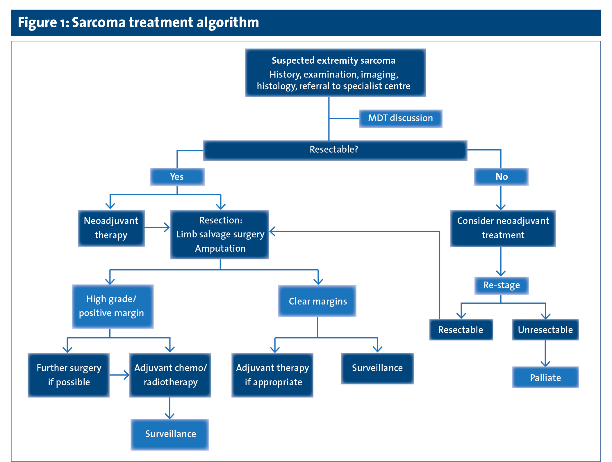
    All suspected sarcomas should be promptly referred to a specialist centre where sarcomas are dealt with frequently (see Figure 1).

     (click to enlarge)

    Soft tissue sarcomas 

    Non-metastatic soft tissue sarcomas are most commonly managed by a combination of radiotherapy and surgery. Radiotherapy may be neoadjuvant (prior to surgical resection) to help reduce the tumour load prior to resection, or adjuvant (after surgical resection). 

    Surgery usually consists of wide excision, with limb salvage. However, margins may occasionally be compromised to preserve function and amputation is sometimes necessary. Chemotherapy is usually reserved for metastatic disease and some paediatric soft tissue sarcomas (mainly rhabdomyosarcoma). 

    Bone sarcomas 

    Management of malignant bone sarcomas depends on the type. Osteogenic sarcomas are managed by a combination of chemotherapy, radiotherapy and surgery. Compared with surgery alone, multi-modal treatment of high-grade non-metastatic osteogenic sarcoma increases disease-free survival from only 10-20% to an excess of 70%. The tumour histological response to pre-operative chemotherapy is an important prognostic factor.9,10 Doxorubicin, cisplatin, methotrexate, ifosfamide and etoposide all have anti-tumour activity in osteogenic sarcoma.11,12,13

    Generally speaking, there is no definite indication for radiotherapy with osteogenic sarcomas but it can be used to extend progression-free interval in sarcomas in anatomic locations where possibility of complete surgical resection is limited. Surgery can be either a limb salvage procedure or amputation, depending on the anatomic site and its relationship to surrounding vital structures.

    Similar to osteogenic sarcoma, treatment of chondrosarcoma is multi-modal. Most chondrosarcomas are low grade, locally aggressive, non-metastasising tumours.14 Grade 1 central chondrosarcomas in long bones of extremities can be treated by curettage with or without adjuvant (eg. cement) with excellent prognosis. Low-grade peripheral chondrosarcomas require surgical excision with a covering of normal tissue over it. Higher-grade chondrosarcomas and all chondrosarcomas of the axial skeleton or pelvis should be surgically excised with wide margins. 

    Contemporary literature suggests that mesenchymal chondrosarcoma may be chemotherapy-sensitive and might benefit from adjuvant or neoadjuvant chemotherapy.15 The value of radiotherapy in chondrosarcoma is limited, but may be indicated in selected cases or for palliation.

    For Ewing’s sarcoma, complete surgical excision (if possible) is considered the best modality of local control, given the relatively high risk of local recurrence when radiotherapy is used alone. Chemotherapy has an important role in both adjuvant and neoadjuvant settings and has significantly improved outcomes. Agents considered most active include doxorubicin, cyclophosphamide, ifosfamide, vincristine, dactinomycin and etoposide.16,17 Radiotherapy alone should be applied if complete surgical excision is not possible.18 Adjuvant radiotherapy should be given in the case of inadequate surgical margins or when histological response in the surgical specimen is poor (> 10% viable tumour cells).

    Recovery and surveillance 

    Resection of either a soft tissue or bone sarcoma is potentially a very invasive procedure with associated emotional and physical disability and overall reduction in quality of life. Predictors of worse functional outcome include large tumour size, high-grade, deep tumours, sacrifice of a major motor nerve and resection of bone.19 To determine the impact of surgery on a patient, functional outcome scores have been developed including the Toronto Extremity Salvage Score (TESS)20 and the Musculoskeletal Tumour Society (MSTS) scoring system.21

    Recurrence is perhaps the most devastating outcome for both the patient and the surgeon. The vast majority of local recurrence and lung metastases occur within the first two years following definitive treatment. Therefore, high-risk patients are followed up every three months for the first two years for clinical examination and a chest radiograph or CT scan.

    Jessica Ryan is an orthopaedic senior house officer and Yasser Aljabi is an orthopaedic registrar at the Department of Orthopaedic Surgery, St Vincent’s University Hospital (SVUH), Dublin, and Gary O’Toole is a consultant orthopaedic surgeon at the Departments of Orthopaedic Surgery in SVUH, Cappagh Hospital, Dublin, and the Beacon Hospital, Sandyford

    References 

    1. Krush AJ. The classification of tumors by John Abernethy early in the nineteenth century. Transactions of the Nebraska Academy of Sciences and Affiliated Societies. 290. 1980
    2. Fletcher CD. The evolving classification of soft tissue tumours – an update based on the new 2013 WHO classification. Histopathology 2014; 64(1): 2-11
    3. Miller RW, Young JL Jr, Novakovic B. Childhood cancer. Cancer 1995; 75(1 Suppl): 395-405
    4. Sinha S, Peach AH. Diagnosis and management of soft tissue sarcoma. BMJ (Clinical research ed) 2010; 341: c7170
    5. Mankin HJ, Mankin CJ, Simon MA. The hazards of the biopsy, revisited. Members of the Musculoskeletal Tumor Society. The Journal of Bone and Joint Surgery American volume 1996; 78(5): 656-63
    6. Rehders A, Stoecklein NH, Poremba C, et al. Re-excision of soft tissue sarcoma: sufficient local control but increased rate of metastasis. World Journal of Surgery 2009; 33(12): 2599-605
    7. Enneking WF, Spanier SS, Goodman MA. A system for the surgical staging of musculoskeletal sarcoma. Clinical Orthopaedics and Related Research 1980; (153):106-20
    8. Edge SB, Compton CC. The American Joint Committee on Cancer: the 7th edition of the AJCC cancer staging manual and the future of TNM. Annals of Surgical Oncology 2010; 17(6): 1471-4
    9. Bielack SS, Kempf-Bielack B, Delling G, et al. Prognostic factors in high-grade osteosarcoma of the extremities or trunk: An analysis of 1,702 patients treated on neoadjuvant co-operative osteosarcoma study group protocols. Journal of Clinical Oncology 2002; 20(3): 776-90
    10. Picci P, Sangiorgi L, Rougraff BT, et al. Relationship of chemotherapy-induced necrosis and surgical margins to local recurrence in osteosarcoma. Journal of Clinical Oncology 1994; 12(12): 2699-705
    11. Lewis IJ, Nooij MA, Whelan J, et al. Improvement in histologic response but not survival in osteosarcoma patients treated with intensified chemotherapy: a randomized phase III trial of the European Osteosarcoma Intergroup. Journal of the National Cancer Institute 2007; 99(2): 112-28
    12. Ferrari S, Smeland S, Mercuri M, et al. Neoadjuvant chemotherapy with high-dose ifosfamide, high-dose methotrexate, cisplatin, and doxorubicin for patients with localized osteosarcoma of the extremity: A joint study by the Italian and Scandinavian Sarcoma Groups. Journal of Clinical Oncology 2005; 23(34): 8845-52
    13. Carrle D, Bielack SS. Current strategies of chemotherapy in osteosarcoma. International Orthopaedics 2006; 30(6): 445-51
    14. Eefting D, Schrage YM, Geirnaerdt MJ, et al. Assessment of interobserver variability and histologic parameters to improve reliability in classification and grading of central cartilaginous tumors. The American Journal of Surgical Pathology 2009; 33(1): 50-7
    15. Cesari M, Bertoni F, Bacchini P, et al. Mesenchymal chondrosarcoma: An analysis of patients treated at a single institution. Tumori 2007; 93(5): 423-7
    16. Bernstein M, Kovar H, Paulussen M, et al. Ewing’s sarcoma family of tumors: current management. The Oncologist 2006; 11(5): 503-19
    17. Paulussen M, Craft AW, Lewis I, et al. Results of the EICESS-92 Study: Two randomized trials of Ewing’s sarcoma treatment – cyclophosphamide compared with ifosfamide in standard-risk patients and assessment of benefit of etoposide added to standard treatment in high-risk patients. Journal of Clinical Oncology 2008; 26(27): 4385-93
    18. Cotterill SJ, Ahrens S, Paulussen M, et al. Prognostic factors in Ewing’s tumor of bone: Analysis of 975 patients from the European Intergroup Cooperative Ewing’s Sarcoma Study Group. Journal of Clinical Oncology 2000; 18(17): 3108-14
    19. Davis AM, Sennik S, Griffin AM, et al. Predictors of functional outcomes following limb salvage surgery for lower-extremity soft tissue sarcoma. Journal of Surgical Oncology 2000; 73(4): 206-11
    20. Davis AM, Wright JG, Williams JI, et al. Development of a measure of physical function for patients with bone and soft tissue sarcoma. Quality of Life Research 1996; 5(5): 508-16
    21. Enneking WF, Dunham W, Gebhardt MC, et al. A system for the functional evaluation of reconstructive procedures after surgical treatment of tumors of the musculoskeletal system. Clinical Orthopaedics and Related Research 1993; (286): 241-6
    © Medmedia Publications/Cancer Professional 2018