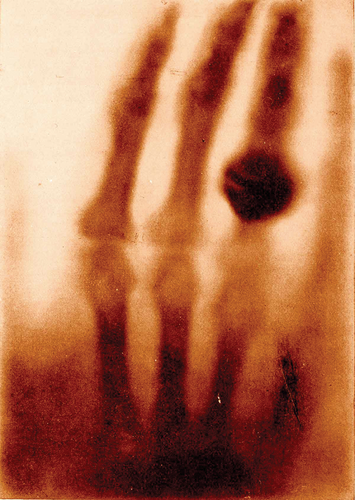
A German physicist experimenting with his newly discovered invisible ray chanced to pass his hand through the beam. On the adjacent screen, the flesh of his hand had seemingly melted away, becoming a ghostly shadow through which only the bones were visible. Had Prof Wilhelm Röntgen any inkling in that heart-stopping moment on November 8, 1895, that he had just stumbled upon one of the most important discoveries in medical history?
Prof Röntgen delivered a paper detailing his findings in December of that year, admitting that the precise nature of these new rays was unknown to him, and calling them x-rays, “for the sake of brevity,” as ‘x’ is the mathematical symbol for the unknown. This new ray, unlike any others known at that time, was able to penetrate solid objects, even the thick walls of his laboratory, but it was the skeletal hand that captured the imagination of the public and of doctors, who immediately recognised that this discovery could transform medical practice forever. The science of medical radiology was born.
Evolution
More than a century later, medical imaging has evolved into a vastly more sophisticated art form, yet it is still based on the principles that different parts of the body absorb a beam of x-ray according to their density, producing an image in which internal structures can be identified as well as any abnormalities indicative of injury and disease.
X-rays are electromagnetic waves, like light waves but with a wavelength about one thousand times smaller. Because of this very short wavelength, x-rays can easily penetrate low-density material, such as flesh, but are reflected or absorbed by high-density material, such as bone. The picture made by an x-ray machine shows denser materials as dark areas.
In a chest x-ray image, the calcium density of the spine and ribs blocks most x-rays, leaving white areas on a film. The water densities of the stomach and liver appear grey as they block less of the x-ray beam than bones. The fat density of muscles is less than that of the water so they look only slightly darker; finally, the air spaces in the lungs allow penetration of most of the x-ray beam and look almost black on the films.
While it sounds straightforward enough, x-ray films require a highly trained eye to recognise the distinction between superimposed ‘shadows’ of varying shades from back to white, interpret the results and make a diagnosis.
‘A million indications’
“Plain film radiograph, or x-ray, is the oldest, simplest and most common form of medical imaging. It’s basically a two-dimensional representation of a three-dimensional structure,” explained Dr Eoin Kavanagh, a consultant radiologist at the Mater Misericordiae Hospital and Cappagh National Orthopaedic Hospital, and senior lecturer at UCD School of Medicine and Medical Science.
“There are a million indications for x-rays: does the patient have a broken bone? Is there a foreign object in the body? Do they have a chest infection? The list goes on and on. Pretty much every patient who enters the hospital system will get some form of x-ray examination performed.
“This is the most standard of all our procedures, where you shine the x-ray beam through the patient and onto an image detector and then we interpret the resulting film. I would say in the Mater Hospital we do a couple of hundred x-ray examinations every day. In Cappagh, where I work also, we do many thousands every month,” he added.
Besides diagnostic applications, interventional radiologists such as Dr Kavanagh use x-ray and other imaging techniques to guide ‘real-time’ procedures, such as needle biopsies. “This afternoon, for example, I’ll see about 10 patients for joint injections during which I’ll use x-ray guidance that we call fluoroscopy, which is low-dose x-ray, to guide the needle tip into various joints. Not all of our image-guided procedures involve x-rays; we use ultrasound and some MRI also.”
Early interventions
Thomas Edison is credited with developing the fluoroscope in 1896, a calcium tungstate-coated screen that glowed when x-rays hit it, allowing direct viewing of any part of the anatomy. At that time, x-ray techniques were advancing apace but it soon became apparent that, while low doses of x-ray appeared to have a good effect on many skin diseases, repeated exposure to high-dose x-rays – a source of radiation – was dangerous, and could prove deadly. Edison dropped x-ray research around 1903 after Clarence Dally, his assistant in x-ray research, died of extreme and repeated x-ray exposure.
In the early 20th century, the problem of looking within body structures was finally addressed when it was found that liquids opaque to x-rays could be ingested or placed within a patient, which allowed the viewing of structures, blockages, ulcers, cancers and other defects. One such material was the common mineral barium sulphate, which could be ground up and swallowed to outline the oesophagus, stomach, and small intestine.It could also be inserted as an enema to visualise the large intestine. It is still used to this day, most often in imaging of the gastrointestinal tract during what is colloquially known as a ‘barium meal’. Other radio-contrast agents were also developed that could be used with the kidneys, the brain and spinal canal, the circulatory system and the lungs.
In the 1970s, diagnostic radiology made a huge leap into cross-sectional imaging when medical engineers added computers to the equation, developing computerised axial tomography (CAT or CT scanning), in which multiple cross-sectional x-ray views are combined by a computer to create three-dimensional images of the body’s internal structures.
Detail from the first-ever medical x-ray, the hand of Mrs Wilhelm Röntgen, 1895(click to enlarge)
More recent developments
Around the same time that CT scanning was becoming a practical tool, a less harmful and more revealing imaging technology appeared on the scene that did not rely on x-rays: magnetic resonance imaging (MRI), which uses powerful magnets and radio waves to create pictures of the body.
While x-rays can show only the anatomical structures and nothing else, the advent of positron emission tomography (PET) in the mid-70s allowed doctors to study metabolic activity using a short-lived radioactive substance to produce three-dimensional coloured images of how those substances are functioning within the body.
“More recent advances in x-ray have been in areas like developing systems which are filmless, digital transfer of images, reading them on monitors and so on. In terms of future developments in x-ray, it is probably a technology which has evolved as far as it will go,” said Dr Kavanagh. “In terms of radiology, however, we’re looking at fusing PET scanning with MRI; that’s going to be a massive area of development. A PET-MRI scan is where a patient has a PET and a MRI scan at the same time so that you can identify ‘hot spots’ and then map them onto the MRI scan. That’s a very attractive option because the CT scan with current PET-CT scanners comes with quite a high radiation dose, and it would be better to avoid that.”
The future of medical imaging
In this age of personalised medicine, molecular imaging is delivering on the promise of providing patient-specific information that allows treatment to be tailored to the exact biological characteristics of both the disease and the patient.
“Imaging can be anatomic, which is where we’re looking inside the body purely anatomically, or it can be functional. With functioning imaging or molecular imaging we can actually see molecular processes that are happening in the body.
“What are the functions of the underlying cells? What cells are different in this patient’s body, what is dividing at the wrong rate? This lump in the lung, has it got a high metabolic rate? Could it be a tumour? Do we need to biopsy it? Molecular imaging can help us to answer those questions,” said Dr Kavanagh. “X-ray has an important place in our arsenal when we are diagnosing or following up a patient but we have far more detailed imaging techniques now. It would be fair to say that the discovery of x-ray really got the ball rolling for radiology more than 100 years ago, and it hasn’t stopped since.”
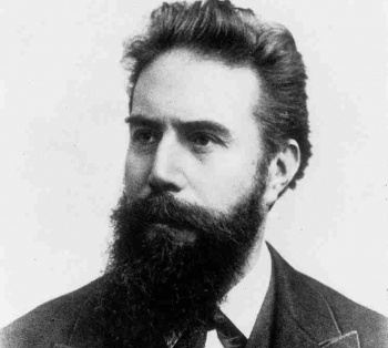 Wilhelm Röntgen(click to enlarge)