ENDOCRINOLOGY

Fibrous dysplasia: challenges in diagnosis and treatment

Fibrous dysplasia can be associated with several extra-skeletal manifestations that can complicate its diagnosis and treatment

Dr Malachi McKenna, Consultant Endocrinologist, St Michael's Hospital, St Vincent's Care Group, Dun Laoghaire, Co Dublin and Dr Anne McGowan, Specialist Registrar, St Vincent's University Hospital, Dublin

February 3, 2016

Article
Similar articles
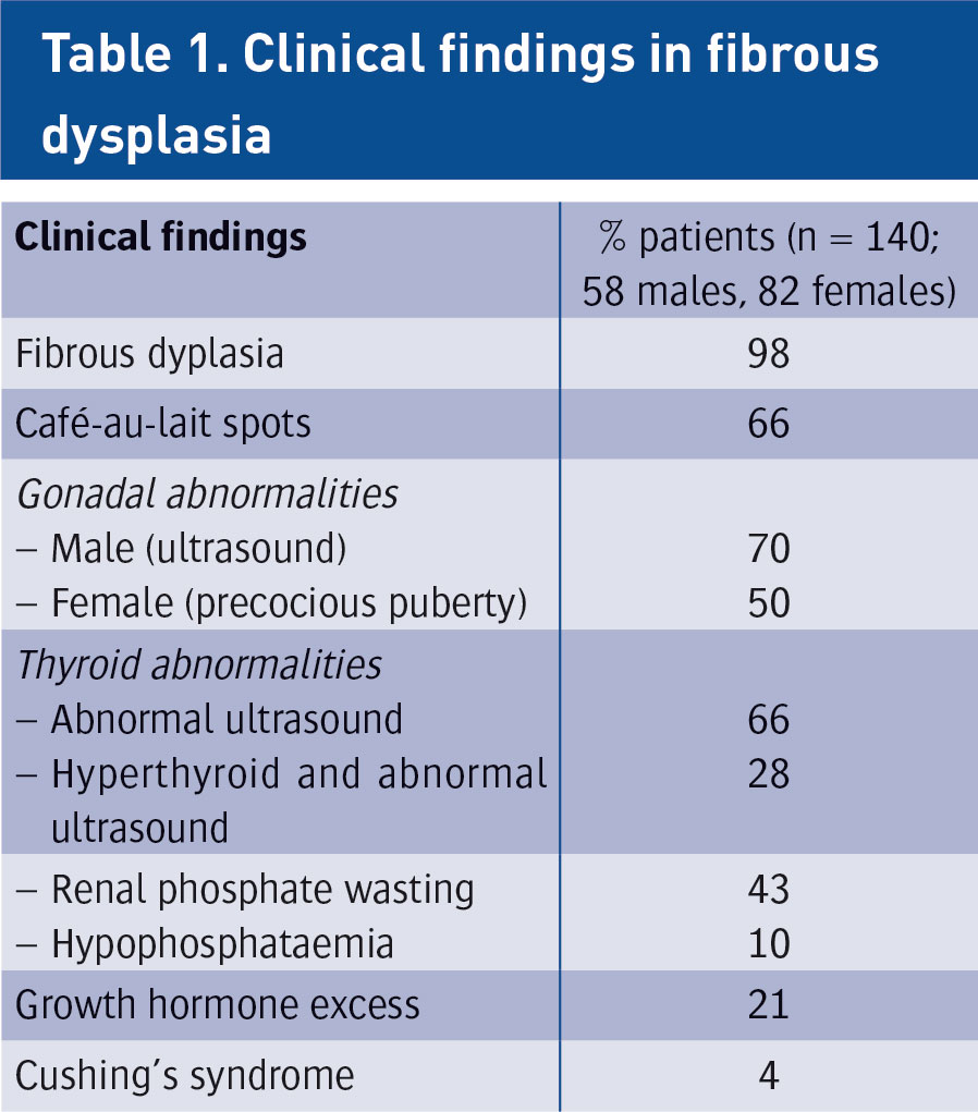
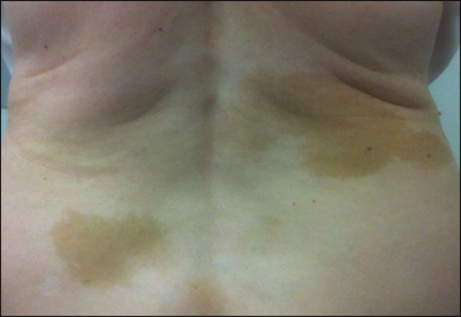
  • Fibrous dysplasia (FD) is an uncommon and debilitating skeletal disorder that typically presents to orthopaedic surgeons in Ireland for diagnosis and management. It can result in deformity, fracture, functional impairment and pain.1 Disease severity is highly variable and may affect one bone (monostotic FD) or many bones (polyostotic FD). Further complicating the diagnosis and treatment of FD is that it can be associated with a wide array of extra-skeletal manifestations. Common extra-skeletal manifestations include café-au-lait macules and hyperfunctioning endocrinopathies including hyperthyroidism, hypercortisolism, precocious puberty and growth hormone excess. One of the most common extra-skeletal manifestations of FD is a renal tubulopathy, which includes renal phosphate wasting. When extra-skeletal manifestations occur, the disease is referred to as McCune-Albright syndrome (MAS). The high degree of phenotypic variability makes the diagnosis and treatment of FD/MAS challenging.

    Pathogenesis

    FD arises from activating mutations in the GNAS gene, which encodes the alpha-subunit of the Gs stimulatory protein (Gs-alpha). Mutations arise postzygotically, leading to mosaic disease with a wide clinical variability. The stimulatory downstream effects of constitutively activated Gs-alpha results in increased adenylyl cyclase activity and inappropriate intracellular cyclic adenosine monophosphate (cAMP) production. Occurring in bone, this results in proliferation of undifferentiated bone marrow stromal cells resulting in marrow fibrosis and increased osteoclastogenesis. Bone trabeculae are abnormal in shape and biochemical composition, and in many cases are severely under-mineralised and abnormally compliant. Elevated serum levels of fibroblast growth factor 23 (FGF23), a phosphate-regulating hormone produced by highly activated osteoblastic cells in FD, are thought to result in the renal phosphate wasting associated with the extra-skeletal manifestations of FD. The pathological effects of Gs-alpha mutations in bone are more pronounced during phases of rapid bone growth, accounting for the fact that childhood and adolescence are when the disease most commonly presents.

    Clinical features

    The most common presenting features include pain, limp and fractures. Presentation before the age of five typically heralds more severe disease, likely to lead to greater morbidity. In general, children are less likely to complain of pain per se and may report stiffness or tiredness. In adults, presentation with pain is more common, especially in the long bones, ribs and craniofacial bones. Spinal and pelvic lesions are typically less painful. Pathological fractures or stress fractures in weight bearing limb bones are the main cause of morbidity. Deformities in bones are due to expansion and abnormal compliance of FD-affected bones and local complications such as cyst formation. 

    Diagnosis is usually made on clinical grounds, based on radiographic appearances and location. Areas of skeletal involvement are established early with 90% of craniofacial lesions evident by five years of age and 75% of all FD lesions established by 15 years of age. Although any area of the skeleton can be affected, common sites include the skull base and proximal femora. Skull base disease may result in facial asymmetry and, rarely, cranial nerve involvement. Femoral disease may present with a classic shepherd’s crook (coxa vara) deformity. Radiographically, these sites appear different. Craniofacial FD has a sclerotic appearance on plain film and ‘ground glass’ on CT; and over time they become more inhomogeneous and lytic on CT. Long bone disease typically displays a homogenous ‘ground glass’ appearance on plain film and becomes more sclerotic as the disease becomes quiescent with age (see Figure 1). The diagnosis can be supported by histological examination of biopsy material or mutation testing of the affected area. The majority of skeletal disease depending on the site is clinically manifest by three to 10 years of age. Markers of bone turnover are usually increased.

    Figure 1. Radiographic findings on fibrous dysplasia in the femur
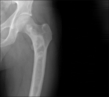
    Figure 1. Radiographic findings on fibrous dysplasia in the femur(click to enlarge)

    At presentation, extra-skeletal manifestations should be identified. Certain conditions, ie. hypophosphataemia or hyperthyroidism, can significantly worsen the clinical course of FD. Thorough phenotyping at presentation allows for identification of all affected tissues but also reassures the patient and their family. The prevalence of extra-skeletal manifestations in patients with FD/MAS are presented in Table 1 from a cohort in the National Institute of Health and likely represents a more severely affected group than that seen in the general population. In the same cohort, a number of other findings associated with the disease have been described. These include gastrointestinal findings (7%); (reflux, hepatitis, pancreatitis and polyps), cardiac findings (4%) including tachycardia, cancer (4%), and neuropsychiatric conditions (9%). Café-au-lait spots that are present in MAS are typically the first manifestation of the disease; they may present at or shortly after birth, and can be used as a clue to an early diagnosis (Figure 2). There is no association between the size of the café-au-lait spots and disease severity. 

     (click to enlarge)

    Figure 2. Café-au-lait manifestation of fibrous dysplasia
    Figure 2. Café-au-lait manifestation of fibrous dysplasia(click to enlarge)

    Precocious puberty is one of the defining manifestations of MAS. It is more common in girls than boys and presents in girls with vaginal bleeding, usually due to a haemorrhagic ovarian cyst. On examination, breast development is often noted to be increased at Tanner stage II-III. Biochemical diagnosis relies on elevated estradiol levels in the setting of suppressed gonadotropins. A pelvic ultrasound typically reveals a large unilateral ovarian cyst, which may have mixed solid and cystic components. The unilateral presentation of a cyst contrasts with the bilateral presentation in central precocious puberty due to hypothalamic-pituitary-gonadal axis activation. Precocious puberty in boys is very rare and when present is more likely to be indolent and subtle. 

    Thyroid disease is very common in MAS when assessed by ultrasound. Typically, the thyroid is enlarged and multinodular and it is important to check for TSH suppression, especially in children as untreated hyperthyroidism is associated with advanced bone age, which can be a problem, especially in the context of precocious puberty. Growth hormone excess is always associated with skull base FD. It is an important diagnosis to make because it is associated with greater morbidity in the craniofacial region. 

    Initially, it was reported in 1968 that rickets was associated with FD. It is now known that a circulating phosphaturic hormone, similar to that observed in the inherited form of rickets, is the cause. Overproduction of FGF23 by the FD affected bone results in renal phosphate wasting. The concentration of FGF23 correlates with disease severity, so the higher the FGF23, the greater degree of renal phosphate wasting and the lower the serum phosphorus. The clinical manifestations of low serum phosphorus are earlier age of first fractures and more fractures and pain. 

    Management and treatment

    The diagnosis of FD depends on clinical, radiographic and histopathological features. The extent of the bone involvement is best determined with a whole body bone scan. Mutational analysis can be performed in order to distinguish it from other forms of fibro-osseous lesions of the skeleton. Disease of the proximal femur may be amenable to prophylactic intramedullary nailing to prevent serious deformities and limb length discrepancy. Surgery is not advocated in craniofacial involvement unless there is hearing or visual loss documented. 

    Antiresorptive therapy with bisphosphonates has been advocated based on observational data due to high rates of bone turnover. Early studies were promising, demonstrating improvement in pain, decrease in bone turnover markers and improved radiographic features of FD. Longer term studies reported similar results. More recently, a double-blind placebo-controlled randomised study of alendronate over two years in FD was reported.2 Clinical data was collected on 24 adults and 16 children and results were available for 35 individuals. Overall, alendronate led to an improvement in areal bone mineral density but no improvement in pain or functional parameters. 

    We favour treatment with intravenous zoledronic acid once yearly up to a maximum of three years based on response both clinically and to bone turnover markers. 

    It is evident that there is a spectrum of extra-skeletal manifestations in FD/MAS but many of these conditions once identified are manageable and potentially treatable in many of the conditions listed. 

    References
    1. Collins MT, Riminucci M, Bianco P. Fibrous Dysplasia. In: Primer on the Metabolic Bone Diseases and Disorders of Mineral Metabolism Eight ed. Rosen CJ, editor: John Wiley and Sons Inc; 2013: 786-793
    2. Boyce AM, Kelly MH, Brillante BA, Kushner H et al. A randomized, double blind, placebo-controlled trial of alendronate treatment for fibrous dysplasia of bone. J Clin Endocrinol Metab. 2014 Nov;99(11):4133-40
    © Medmedia Publications/Hospital Doctor of Ireland 2016