RHEUMATOLOGY

CANCER

PAIN

WOMEN’S HEALTH

SURGERY

Sacroiliac joint dysfunction – a common cause of low back pain

The most common cause of sacroiliac joint pain is idiopathic and may occur acutely or insidiously with cumulative trauma

Mr Michael Leonard, Consultant Orthopaedic Surgeon, National Centre for Pelvic and Acetabular Surgery, Tallaght Hospital, Dublin

May 1, 2015

Article
Similar articles
  • The sacroiliac joint (SIJ) is the largest axial joint in the musculoskeletal system and its primary responsibility is to transfer the weight of the upper body to the lower extremities. In the past this joint has been largely ignored as a pain generator, however, recent research has reported that the sacroiliac complex may be the source of pain in 15-25% of all individuals who present with low back problems.1 To avoid unnecessary lumbar spine surgery, SIJ disorders should be strongly considered in low back pain diagnosis.2

    Anatomy 

    The sacroiliac joints are composed of the sacrum wedged between the ilia. It is the largest axial joint in the human body, with a surface area of approximately 17.5cm2. The morphology of the SIJ not only changes with age but it also varies greatly in size, shape and contour from side to side as well as between individuals. 

    The surface of the joint is flat until after puberty. By age 30, bony ridges are present on the articular surface of the ilium. With time the synovial articular surfaces erode and the synovial cleft narrows. The joint is supported by numerous ligaments, muscle and fascia.

    Biomechanics

    The SIJ functions as a shock absorber, forms a base for the spinal axis, transmits and dissipates upper trunk loads, and facilitates childbirth. Motion in the SIJ is limited to small amounts of rotation and translation. 

    Compression of the sacrum with weight bearing creates a ‘keystone in an arch’ effect: the greater the force through the sacrum, the greater the resistance. 

    Pathology

    The most common cause of SIJ pain is idiopathic and may occur acutely or insidiously with cumulative trauma. Idiopathic SIJ pain may result from a change in the joint’s position or mechanics. Other potential causes include:

    • Trauma
    • Osteoarthritis
    • Infection
    • Inflammatory conditions (sacroiliitis is present in virtually 100% of individuals with ankylosing spondylitis)
    • Metabolic conditions (for example, gout and renal osteodystrophy)
    • Malignancy – metastases to the pelvis account for 40% of all osseous metastasis, second only to those of the spine and iatrogenic, for example, post bone graft harvesting, lumbar spine fusion).

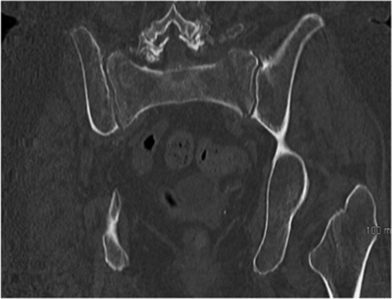
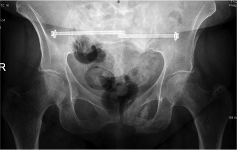
    Insufficiency sacral fractures in the older osteoporotic patient may occur with little or no trauma, and can be difficult to visualise on plain radiographs. Stress fractures have been reported in athletes due to repetitive microtrauma caused by impact loading over long periods of frequent physical exertion. 

    The third trimester of pregnancy results in hypermobility of the sacroiliac joint because of increased levels of oestrogen and relaxin that cause the soft tissues supporting the joint to loosen. This laxity may predispose the sacroiliac joint ligaments to painful sprain. The mechanical trauma of childbirth also may cause joint pain. 

    Diagnosis

    History: Patients with pain originating from the SIJ may complain of a number of different symptoms including:

    • Low back pain
    • Lower extremity pain (numbness, tingling, weakness – usually above the knee) 
    • Buttock pain
    • Hip or groin pain. 

    Functional symptoms, which may be more specific in identifying the SIJ as the source of pain, include:

    • Unilateral leg instability (buckling, giving way) 
    • Disturbed sleep patterns
    • Disturbed sitting patterns (unable to sit for long periods on one side, such as when driving)
    • Transitional symptoms (pain going from sitting to standing or supine to standing) 
    • Pain on the affected side with stair use.

    Examination

    The clinical examination begins with excluding the lumbar spine and hip joint as the main sources of pain with negative screening exams. The following are then performed to isolate the SIJ as the primary source of pain: 

    Fortin test – while standing, have the patient point with one finger to the location of pain, this test is positive if the patient points on two occasions to the area directly medial and inferior to the posterior superior iliac spine (PSIS) (Figure 1)

    Figure 1. Fortin’s finger test
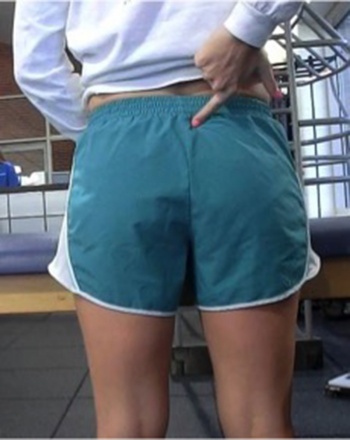
    Figure 1. Fortin’s finger test(click to enlarge)

    A single leg stance test: this may induce pain on the supporting side. In more severe cases the patient may be unable or unwilling to perform this test due to pain or perceived instability

    Deep palpation within the SIJ sulcus (medial and inferior to the PSIS) can isolate SIJ tenderness

    Posterior pelvic pain on active straight leg raise in the supine position

    It is my practice to then perform a thigh thrust (hip flexed to 90 with compression along the long axis of the femur) and a flexion abduction external rotation (FABER) test. Both of these examinations are performed on the affected side with the patient lying on their back while stabilising the contralateral haemipelvis

    An SIJ compression test is then performed with the patient lying on their unaffected side with direct compression applied to the affected SIJ

    Plain film pelvic radiographs and inflammatory markers should be ordered as part of routine work up for SIJ pathology. Advanced imaging may be required depending on the patients history and exam findings

    If positive clinical findings are identified, an image guided SIJ injection that results in a significant improvement in the patient’s symptoms confirms the diagnosis.

    Treatment

    Following diagnosis initial treatment consists of addressing any underlying cause (if present) along with a combination of non-steroidal anti-inflammatories (NSAIDs), muscle relaxants and physiotherapy. 

    A specific physical therapy programme, focusing on decreasing pain and improving function, balance, core stability and correcting lumbopelvic-hip mechanics, is required. Correcting soft-tissue irritability is important, as is establishing a prevention programme and retraining optimal movement patterns through dynamic postural control. In particular, certain muscles should be specifically strengthened. These include the hamstrings, gluteus maximus and medius, piriformis, erector spinae, latissimus dorsi and the iliacus muscles. 

    These measures can if necessary, as dictated by the patients’ symptoms and response to therapy, be combined with intermittent SIJ injections. The injections are performed as day cases with dynamic image guidance.

    Most cases of idiopathic sacroiliac joint/complex pain can be adequately managed with the above modalities, however, due to the wide range of medications and physical therapy treatments available, there is no clear evidence of any one type’s long-term efficacy. In cases that fail to respond to an adequate course (minimum three to six months) of the above, consider operative intervention, specifically fusion of the SIJ. Fusion of the SIJ is now commonly performed percutaneously with the aid of dynamic intra-operative imaging. 

    Reported outcomes of this procedure have been encouraging with up to 88% of patients obtaining substantial clinical benefit and patient satisfaction achieved at 12 months being maintained for five years in 82% of patients post-fusion.3

    Case study 

    A 68-year-old female with a history of chronic low back pain was referred for an opinion. She had undergone extensive investigation of her lumbar spine, low back physiotherapy and analgesia. 

    Figure 2a. Coronal pelvic CT of 68-year-old female with chronic ‘low back pain’ demonstrating bilateral sacral insufficiency fractures
    Figure 2a. Coronal pelvic CT of 68-year-old female with chronic ‘low back pain’ demonstrating bilateral sacral insufficiency fractures(click to enlarge)

    Figure 2b. Plain film pelvic x-ray six months post percutaneous fixation of bilateral sacral insufficiency fractures
    Figure 2b. Plain film pelvic x-ray six months post percutaneous fixation of bilateral sacral insufficiency fractures(click to enlarge)

    Clinical examination was positive for pain emanating from her sacroiliac complex. Plain film pelvic radiographs and CT scan (Figure 2a) demonstrated bilateral sacral insufficiency fractures. Percutaneous bilateral sacroiliac screws were inserted and her fractures went on to heal (Figure 2b) with a significant improvement in her overall symptoms.

    References

    1. Cohen, Steven P. Sacroiliac joint pain: a comprehensive review of anatomy, diagnosis, and treatment. Anesth Analg 2005; 101:1440-1453
    2. Weksler N, Velan GJ et al. The role of sacroiliac (SI) joint dysfunction in the genesis of low back pain: the obvious is not always right. Arch Orthop Trauma Surg 2007; 10(127): 858-888
    3. Rudolf L, Capobianco R. Five-year clinical and radiographic outcomes after minimally invasive sacroiliac joint fusion using triangular implants. Open Orthop J 2014; 8:375-83
    © Medmedia Publications/Hospital Doctor of Ireland 2015