DIABETES
Saving a toe with integrated care
The initial recommendation to amputate an ulcerated toe was avoided thanks to an integrated care team's holistic approach to management
June 1, 2017
-
Ulcerations and foot injuries are major causes of lower extremity amputations in patients with diabetes. Peripheral sensory neuropathy, often with an autonomic or motor component, is the primary factor that leads to foot ulceration in patients with diabetes when combined with unperceived trauma or moderate repetitive stress.
It has been shown that a triad of neuropathy in conjunction with minor foot trauma and foot deformity play a causal role in more than 60% of diabetic foot ulcers.1 Neuropathic disease leads to degeneration of nerves and results in loss of cutaneous sensation.2 The resulting sensory deficit means that if the skin is subjected to continuous trauma – eg. by friction from wearing ill-fitting shoes – damage and finally a break in the skin may occur, all of which goes unfelt and therefore unnoticed by the patient.3
High incidence of infection
There is a relatively high incidence of infection in the wounds of patients with diabetes; however identifying infection in diabetic foot ulcers can prove problematic. The clinical signs and symptoms normally associated with wound infection are frequently less obvious or even absent owing to diabetes-associated complications such as ischaemia, neuropathy and immunological impairment.
Once wound infection has been correctly identified careful management is required in order to arrest wound deterioration which could ultimately result in outcomes such as amputation. Local management of diaebtic foot ulcers will therefore often necessitate the use of antimicrobial dressings to reduce the bacterial burden within the wound bed and enable the wound to progress toward healing.
In addition to the effective treatment and ideally prevention of infection, the management of diabetic foot ulcers must adequately address the multiple factors that will influence healing of the ulcer. A holistic strategy should therefore encompass at least the following:
• Regulation of serum glucose
• Wound debridement
• Pressure off-loading
• Effective dressing selection to optimise the local wound environment.4,5
Achieving the latter can be problematic since many diabetic foot ulcers are in anatomical locations that are difficult to dress, so choice of dressings can be limited.
Case presentation
Ms X, a lady in her mid-60s, had been diagnosed with type 2 diabetes seven years previously, but had failed to engage in the management of her diabetes. Ms X had made none of the lifestyle changes that had been recommended to her following her diagnosis and refused to take any of the medications she had been prescribed. She presented to the clinic with a five-week history of self-treating a wound on her right 2nd toe. She reported that the wound had developed as a result of her shoes rubbing. At this time Ms X was feeling very unwell.
Clinical examination
Prior to any treatment a thorough and comprehensive clinical examination was undertaken. This process allowed a preferred diagnosis to be made and ensured that the management approach taken subsequently was based upon a fully informed appreciation of Ms X’s clinical status and hence gave due consideration to all relevant factors likely to influence the wound.
The examination revealed the following:
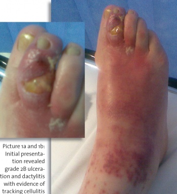
• Grade 2B ulceration6 (infected, non-ischaemic ulcer that penetrates to capsule or bone) to the dorsal aspect of the right 2nd toe overlying the distal inter-phalangeal joint (DIPJ) (see Picture 1a and 1b)
• Wound bed 100% slough
• Dactylitis (inflammation of an entire digit) with evidence of tracking cellulitis (see Picture 1) and the patient was systemically unwell
• Elevated right foot temperature: 38.8°C
• Elevated blood glucose: 15.6mmol/L
• Deep tissue swab: Staphlococcus aureus sensitive to penicillin
• Vascular assessment revealed that the limb was not critically ischaemic: all pedal pulses palpable; Doppler signals biphasic; right foot ankle brachial pressure index 0.91
• Neurological sensory assessment revealed significant peripheral sensory deficit with Ms X being unable to detect either large fibre (light touch and vibration perception) or small fibre (pain and temperature) modalities
• Neurological motor assessment identified clawing of the digits to both feet and an ankle equinus deformity, both suggestive of motor neuropathy
• Neurological autonomic assessment identified clinical features suggestive of autonomic neuropathy: bounding pedal pulses and gross anhydrosis (deficiency or absence of sweating)
• Footwear appraisal: shoes were narrow-fitting dress shoes with shallow toe box providing little room to accommodate Ms X’s clawed digits.
This lack of room coupled with Ms X’s loss of sensation was felt to provide the most likely explanation for the development of the ulcer. Although flat, the shoes had a very hard midsole with thin inflexible insoles and therefore offered minimal shock attenuation.
Initial management
Ms X was immediately admitted to hospital where she remained an inpatient for five days. During her stay, she received intravenous antibiotic therapy to address the wound infection and cellulitis.
Radiological examination identified subluxation of the distal phalanx of the ulcerated toe with an old fracture to the phalanx. The presence of osteomyelitis was not evident on the x-ray.
Ms X was reviewed by the orthopaedic surgical team who recommended amputation of the digit as the best course of action. Ms X was unwilling to give her consent for this procedure and requested that the wound be managed conservatively to see if surgical intervention could be avoided with amputation being performed only as a last resort.
Wound management
Ms X was referred to the podiatry department for wound management following her discharge from hospital. Oral antibiotic therapy with flucloxacillin was continued for a further 14 days post discharge. The wound was sharp debrided to remove a proportion of the slough within the wound bed. Owing to the location of the ulcer there was a need for any dressing used to be both sufficiently thin and conformable to allow the 2nd toe to be enclosed yet without displacing the neighbouring digits.
The patient was also only able to attend clinic appointments weekly due to relying on others for transport, so the dressing applied would need to be capable of being changed only once per week. ACTICOAT Flex 7 was therefore selected as a primary dressing on the basis of its seven-day broad spectrum antimicrobial action and highly conformable nature.
An open toe Darco wound boot was worn to completely off-load the digit. The patient also attended appointments with the dietitian, diabetes nurse specialist and began taking the medication prescribed for the regulation of her blood glucose levels (metformin, gliclazide). The public health nurse was an integral part of management, attending to dressings in the community.
Clinical outcomes
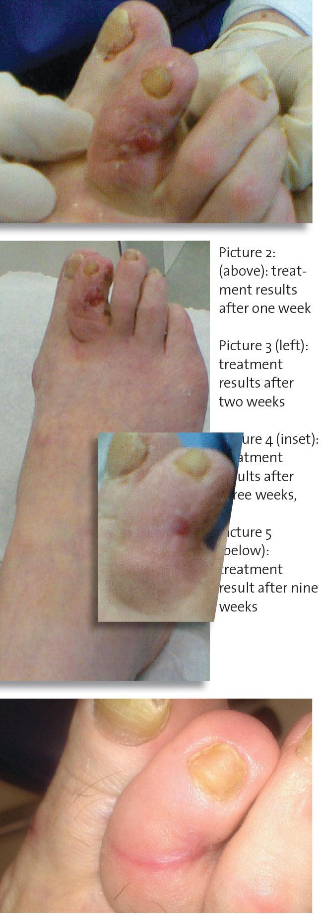
The wound responded well to the treatment regimen of weekly sharp debridement and re-dressing coupled with effective off-loading, with dietary modification and medication to regulate blood glucose levels (see Picture 2).
The evidence of infection, such as the digital oedema, began to resolve and the wound began to make some progress towards healing. The management regimen was continued without modification due to the success it was having and the progress seen initially was maintained in subsequent weeks (see Picture 3).
After three weeks of treatment the wound had fully epithelialised (see Picture 4). Given the poor condition of the wound at initial presentation, achieving epithelialisation after three weeks of treatment was seen as a very successful outcome.
At this point the patient continued to dress the digit with ALLEVYN Adhesive in order to protect the delicate newly formed skin. Ms X was pleased that she was able to do this herself, allowing the frequency of her clinic visits to reduce accordingly.
The integrated care team arranged for further education with the DESMOND (Diabetes Education and Self-Management for Ongoing and Newly Diagnosed) programme delivered in primary care. An orthotist referral was made so that accommodative footwear could be supplied for future ulcer prevention. See Picture 5 for toe after nine weeks.
Conclusion
Following comprehensive assessment, all diabetic foot ulcers should be treated holistically with measures including interventions such as sharp debridement when appropriate, effective dressing regimes, including the selective use of antimicrobial dressings to reduce wound bioburden, adequate off-loading strategies and control of blood glucose.
The wounds of patients with diabetes present a number of clinical challenges to the practitioners who treat them. These challenges are not unique to this patient group but confounding factors, such as neuropathy leading to loss of sensation and physical malformations, may enhance the problems.
The wound location, especially with regard to digit ulcerations, makes application of dressings difficult. Although the evidence presented in this article refers to a specific case, it illustrates how in a real-life situation, the clinical challenges presented by diabetic foot ulceration were successfully overcome via an effective dressing regimen, control of blood glucose and education utilising the integrated care team providing an appropriate holistic management approach.
 (click to enlarge)