The important role of radiology in Irish hospitals
The role of radiologists, who are an integral part of the multidisciplinary team that manages cancer patients, is reviewed
Dr Orla Doody, Radiology Specialist Registrar, Department of Radiology, Tallaght Hospital, Dublin, Dr William C Torreggiani, Consultant Radiologist, Department of Radiology, Tallaght Hospital, Dublin and Dr Terri Hok, Specialist Registrar, Tallaght Hospital, Dublin
Cancer is one of the leading causes of death worldwide. In Ireland, one in three men and one in four women will develop invasive cancer.1 Between 2007 and 2009, 29,745 cases of malignancy were diagnosed in Ireland. In 2007, the mortality from all cancers was 12.6%.1 Medical imaging has evolved to provide improved cancer detection. In addition, radiology is integral to the staging, treatment and ongoing follow-up of patients with cancer.
Screening programmes
The cancer screening programmes in this country utilise imaging in the prevention and early detection of many cancers. Radiological investigations result in the detection of many clinically silent malignancies. BreastCheck – the National Breast Screening Programme – under the auspices of the National Cancer Screening Service (NCSS), offers mammograms to all women aged 50 to 64 years. Since commencing in 2000, the programme has detected 5,017 breast cancers in 368,851 women.2
If colonoscopy is incomplete or unsuccessful in patients who have a positive faecal immunochemical test, computed tomography (CT) colonography will be available as the completion examination through the NCSS colorectal screening programme.3,4 This investigation is also indicated in patients who are medically unfit for colonoscopy. CT colonography offers a minimally invasive examination of the colon to evaluate for colorectal polyps and neoplasm. This examination should be performed and reported by a consultant radiologist with specific training in the technique (see Figure 1).
Figure 1. CT colonography (coronal reformat) demonstrating circumferential mural thickening of the sigmoid colon consistent with a sigmoid malignancy(click to enlarge)
Many radiological investigations involve a radiation dose for the patient. In recent years, imaging technology has improved greatly to minimise the radiation dose to the patient while maintaining the diagnostic value of the study. The use of low-dose spiral CT for lung cancer screening is promising, with a recent National Lung Screening Trial reporting a 20% reduction in lung cancer mortality in high-risk patients when screened with low-dose spiral CT compared to conventional chest x-rays (CXRs).5
Detection and staging
In addition to detection during routine screening, a cancer may be diagnosed incidentally while performing the investigation for another clinical presentation. In many cases imaging is performed to confirm the suspicion in patients with a clinically apparent cancer. A cancer may be detected with plain radiographs, ultrasound, mammography, CT or magnetic resonance imaging (MRI [see Figures 2 and 3]). Imaging provides details on the location of a cancer, the extent of disease and the presence of local or distant complications.
Figure 2. Ultrasound of solid mass in the posterior aspect of the thigh in continuity with the sciatic nerve, consistent with a peripheral nerve sheath tumour (PNST) (click to enlarge)
Figure 3. Corresponding coronal T1 and T2 weighted MRI confirmed ultrasound findings of a PNST of the sciatic nerve. Patient had subsequent staging CT of thorax, abdomen and pelvis(click to enlarge)
Following the detection of a cancer, the role of the radiologist is to stage the disease, typically with a combination of CT, MRI and positron emission tomography (PET) CT. Endoscopic ultrasound may be performed by gastrointestinal radiologists and has a role in the staging of upper gastrointestinal malignancies. Molecular imaging contributes further information regarding the tumour’s metabolism.
PET is typically combined with CT (PET-CT) to provide detail on the metabolic activity of tumours and to detect metastatic disease (see Figure 4). In Ireland, a number of cancers, depending on the stage of the initial cancer on cross-sectional imaging and on the type of malignancy, will have additional staging with PET-CT. The Faculty of Radiologists provides guidelines based on international standards outlining current indications for PET-CT.6 If histological confirmation is required an ultrasound or CT-guided biopsy is often performed by the radiologist.
Figure 4. PET-CT demonstrating avid tracer uptake in a solitary hepatic lesion consistent with a malignancy(click to enlarge)
The extent of a tumour will determine the initial stage of disease and determines the most appropriate treatment plan for a patient. Accurate staging relies on the skill and expertise of the radiologist. Local and distant metastases generally follow a typical pattern depending on the underlying primary diagnosis. These patients are discussed at multidisciplinary team meetings with radiologists, oncologists, physicians, surgeons and pathologists, who together will decide the appropriate treatment pathway for the patient. Radiation oncologists use pre-treatment imaging to decide on an appropriate radiation field to minimise any unnecessary radiation.
Therapeutic options
Interventional radiologists perform many minimally invasive treatment procedures for malignancy. Radiofrequency ablation, trans-arterial embolisation (TAE) and trans-arterial chemoembolisation (TACE) can be utilised in the management of many malignancies, as curative, preoperative or palliative procedures (see Figure 5).7,8,9
Figure 5. Selected images during a trans-arterial embolisation of hepatic metastases. Neovascularity demonstrated from a proximal branch of the right hepatic artery corresponding to a metastatic deposit. Selective coil embolisation performed with post-procedure angiogram demonstrating cessation of blood supply to the tumour(click to enlarge)
Follow-up imaging
Imaging has a major role in monitoring the effectiveness of treatment in patients and allowing treatment to be altered appropriately based on the disease response. The Response Evaluation Criteria in Solid Tumours (RECIST) criteria are the most commonly used criteria defining response, stabilisation or progression, based on tumour and lymph node size. The follow-up imaging of oncology patients requires significant resources within a radiology department. Most commonly, patients undergo a restaging CT with the frequency of scans determined by international guidelines and decided in a multidisciplinary team setting. Ultrasound and MRI may also be utilised depending on the initial malignancy.
References
Cancer in Ireland 2011: Annual report of the National Cancer Registry
Breast Check Programme Report 2010 – 2011
National Cancer Screening Service. Guidelines for quality Assurance in Colorectal Screening
Faculty of Radiologists. Guidelines for use of CT Colonography as Part of the National Colorectal Screening Programme in Ireland
National Lung Screening Trial Research Team, Aberle DR, Adams AM, Berg CD et al. Reduced lung-cancer mortality with low-dose computed tomographic screening. N Engl J Med 2011; 365(5): 395-409
Faculty of Radiologists. PET CT Guidelines
Gervais DA, Arellano RS. Percutaneous tumor ablation for hepatocellular carcinoma. AJR 2011; 197(4): 789-94
Arterially directed therapies for hepatocellular carcinoma. Shah RP, Brown KT, Sofocleous CT. AJR 2011; 197(4): W590-602. Review. PMID: 21940531
Hague J, Tippett R. Endovascular techniques in palliative care. Clin Oncol 2010; 22(9): 771-80
Making cancer visible. The role of imaging in oncology. European Society of Radiology
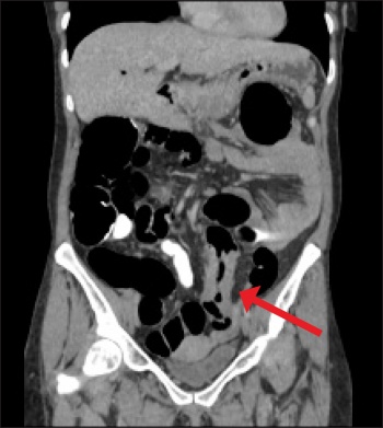 Figure 1. CT colonography (coronal reformat) demonstrating circumferential mural thickening of the sigmoid colon consistent with a sigmoid malignancy(click to enlarge)


