RESPIRATORY

A case of bilateral pulmonary artery hypertrophy

A case of respiratory failure caused by pulmonary artery hypertension with severe hypertrophy on chest x-ray

Dr Christine Newman, Registrar, Mayo University Hospital, Castlebar, Co Mayo and Dr Fionnula Lavin, Consultant Physician, Mayo University Hospital, Castlebar, Co Mayo

October 2, 2017

Article
Similar articles
  • Pulmonary hypertension is an uncommon disorder characterised by mean pulmonary artery pressure ≥25mmHg. It has multiple aetiologies and has a prevalence of 5-50 per one million people.1 In this case we report on a case of respiratory failure caused by pulmonary artery hypertension with severe hypertrophy on chest x-ray. 

    Case

    A 54-year old woman with a history of pulmonary stenosis, pulmonary hypertension (of mixed aetiology), mitral regurgitation and atrial fibrillation presented to the emergency department with chest discomfort and worsening dyspnoea. She was tachycardic, tachypnoeic (at 48 resp/min) with oxygen saturations of 72% on 4L O2. Cardiovascular and respiratory system examination revealed a loud pan-systolic murmur, diffuse wheeze and pitting oedema to the mid-thigh. 

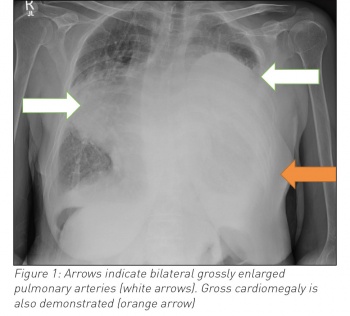
    Blood work showed a neutrophilia, hypochromic anaemia, elevated C-reactive protein and an acidotic type two respiratory failure. A chest radiograph was significant for an opacity in the lower two-thirds of the left lung field and a second opacity in the right mid-zone. Given the woman’s past history the differential included right-sided pneumonia, left-sided pleural effusion and cardiomegaly, or hypertrophied pulmonary arteries.

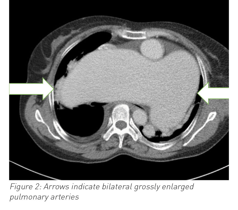
    A computed tomography scan confirmed grossly hypertrophied pulmonary vasculature.   

    Despite intubation, ionotropic and anti-microbial support this woman passed away in the intensive care unit after five days. 

     (click to enlarge)

     (click to enlarge)

    Discussion

    Pulmonary hypertension is a chronic disease with a five year mortality of roughly 57%.2

    The initial investigations for pulmonary hypertension include ECG, chest x-ray, echocardiogram, pulmonary function test and right-heart catheterisation. 

    While chest x-ray can sometimes point towards the underlying cause of pulmonary hypertension (eg. interstitial lung disease), it usually only bears subtle signs. Typically the chest x-ray would show mild enlargement of the central pulmonary arteries or interstitial oedema. Occasionally, an enlarged right atrium can be seen but signs are often subtle and have a low sensitivity and specificity. 

    Despite this roughly 90% of patients will have some abnormality on the chest x-ray by the time a diagnosis of pulmonary hypertension is diagnosed.3

    This case serves to highlight the radiological findings at the extreme end of the spectrum and to raise clinician awareness.

    References

    1. Badesch DB et al. Pulmonary arterial hypertension: baseline characteristics from the REVEAL Registry. Chest 2010;137(2):376
    2. Benza RL et al. An evaluation of long-term survival from time of diagnosis in pulmonary artery hypertension. Chest 2012; 142(20):448
    3. Gaile N et al. Guidelines on diagnosis and treatment of pulmonary arterial hypertension. The Task Force on Diagnosis and Treatment of Pulmonary Arterial Hypertension of the European Society of Cardiology. Eur Heart J 2004; 25(24): 2243-2278
    © Medmedia Publications/Hospital Doctor of Ireland 2017