NEPHROLOGY

PAIN

SURGERY

Aorto-caval fistula: haematuria and acute renal impairment

A case of aorto-caval fistula with haematuria and acute renal impairment

Dr Sheng Chieng Hong, SHO in General Surgery, Portiuncula Hospital, Galway, Dr Louisa Joyce Lim, SHO in the Emergency Department, Portiuncula Hospital, Galway and Mr Eddie Myers, Consultant Colorectal Surgeon, Portiuncula Hospital, Co Galway

November 1, 2013

Article
Similar articles
  • Haematuria and acute renal failure are two well recognised clinical features of aortic aneurysm complicated by aorto-caval fistula (ACF). ACF is a rare condition complicating around 3% of ruptured aortic aneurysms.1 It is difficult to diagnose if clinical suspicion is low. Interestingly enough, it is much more common in men in their 60s.2,3 

    There is no doubt that it is a fatal complication of abdominal aortic aneurysm with a high mortality rate ranging from 21-66.7%.4 In this case report, the authors present an 84-year-old with ACF from a small hospital in the west of Ireland.

    Case report

    The patient self-presented to the emergency department with a six-day history of acute onset of dull back pain and three-day history of gross haematuria. 

    Otherwise, he is a healthy man with no known past medical or surgical history and this being the first admission into hospital. The pain was described to be of a sudden onset, dull in nature and localised to the right flank. He reports no other urinary symptoms, no history of recent trauma, no loss of appetite or weight and no altered bowel habit. 

    Of note, his social history includes a 79-pack-year smoking history since age 14 and is a social drinker. On examination, his vital signs were: blood pressure 90/40mmHg, heart rate 62/min and respiratory rate 22/min. Clinical examination reviewed a persistently low blood pressure. No abdominal masses were found and his chest was clear.

    His blood investigation showed raised urea (29.30mmol/L) and creatinine (330umol/L) indicating acute renal impairment. Haemoglobin was low at 9.7g/dL, WCC raised at 18.5 and a neutrophil count of 16.3 indicating a likely infection. A three-way urinary catheter was inserted for irrigation and more than half a litre of dark red urine and blood clots drained immediately.

    This then raised the suspicion of obstructing renal or ureteric calculi but an urgent plain x-ray of the abdomen did not show any evidence of stones in the urinary tract. 

    Despite vigorous fluid resuscitation and irrigation, the gross haematuria persisted and his blood pressure remained low. An urgent ultrasound of the abdomen was done the following day. A massive aortic aneurysm measuring 6.5cm in diameter (see Figure 1) was revealed.  There was also some fluid collection in the abdomen indicating that there was a possibility that it was a rupturing aortic aneurysm. 

    Figure 1. Abdominal ultrasound scan showing an aortic aneurysm measuring 6.5cm in diameter
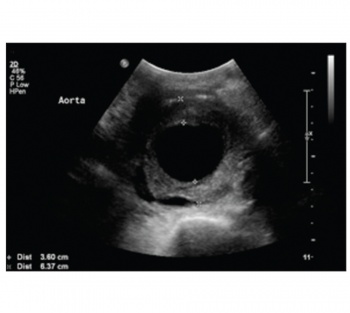
    Figure 1. Abdominal ultrasound scan showing an aortic aneurysm measuring 6.5cm in diameter(click to enlarge)

    Figure 2. CT aortography showing an aortic aneurysm with intramural thrombosis and aorto-caval fistula
    Figure 2. CT aortography showing an aortic aneurysm with intramural thrombosis and aorto-caval fistula(click to enlarge)

    The vascular team in University College Hospital Galway was immediately informed and he was promptly transferred thereafter. CT aortography was performed and confirmed the presence of a leaking aortic aneurysm with an ACF. Figures 2, 3 and 4 show the presence of infrarenal aortic aneurysm with an ACF. He was immediately assessed by the vascular team and an urgent endovascular aneurysm repair (EVAR) and ligation of fistula was done. A total of six stents, including one inferior vena cava stent, were inserted endoscopically via the femoral artery and vein, respectively. 

    He survived the procedure and was subsequently transferred to the intensive care unit (ICU). The gross haematuria resolved within the same day and there was a vast improvement in his renal function. 

    His urea and creatinine came down to 14.0mmol/L and 130umol/L, respectively. He was discharged to a non-acute ward setting at day three post-operatively and was discharged home at day 11 post-operatively. 

    Discussion

    ACFs are commonly caused by enlarging atherosclerotic aorta.6 Trauma such as penetrating abdominal trauma and iatrogenic trauma at lumbar disc surgery can also cause ACF.7 Among the rare causes are mycotic aneurysm, syphilis and connective tissue disorders such as Ehlers-Danlos and Marfan syndrome.6

    Classically, an abdominal aortic aneurysm (AAA) presents with a triad of central abdominal pain which radiates to the back, pulsatile mass in the abdomen and hypotension. However, it is important to recognise that an AAA complicated by ACF could present with only haematuria and acute renal impairment, such as in this case.

    Figure 3. Coronal section of the CT aortography showing the aortic aneurysm with aorto-caval fistula
    Figure 3. Coronal section of the CT aortography showing the aortic aneurysm with aorto-caval fistula(click to enlarge)

    Figure 4. Sagittal section of the CT aortography
    Figure 4. Sagittal section of the CT aortography(click to enlarge)
     

    It has been reported that 7-23% of patients with aorto-caval fistula present with haematuria.3,5

    Salo et al in their 13 years of experience with 11 patients reported that haematuria was present in all of their patients; six had macrohaematuria and five had microhaematuria, showing that, in fact, haematuria should be recognised as a symptom of ACF.8 However, the mechanism for causing haematuria is uncertain. It has been postulated that the fistula causes an increase in pressure in the venous system and presents as dilated veins in the bladder, which bleeds easily.

    Acute renal failure such as that seen in this patient has been described in a few case reports. Brunkwall et al reported that seven of his nine patients studied had acute renal insufficiency with a median 292mmol preoperatively but declined to 86mmol at discharge, concluding that renal function normalised after successful surgery.

    It is said that the renal insufficiency is caused the raised renal venous pressure causing a decrease in the renal arterial perfusion pressure. However, from all of the reported cases of ACF causing acute renal failure, renal function returned to normal after surgery.

    Conventionally, the surgical option for repair of ACF would have resulted in a laparatomy scar, but novel approaches are now an available option to patients. Thus, the patient had a successful EVAR procedure from which he recovered uneventfully.

    References

    1. Baker WH, Sharzer, LA, Ehrenhaft JL. Aorto-caval fistula as a complication of abdominal aortic aneurysms. Surgery 1972; 72: 933
    2. Calligaro KD, Savarese RP, DeLaurentis DA. Unusual aspects of aortovenous fistula associated with ruptured abdominal aortic aneurysms. J Vasc Surg 1990; 12: 586-590
    3. Cinara IS, Davidovic LB, Kostic DM et al. Aorto-caval fistula: a review of eighteen years experience. Acta Chir Belg 2005; 105: 616-620
    4. Alexander JJ, Imbembo AL. Aorto-vena cava fistula. Surgery 1989; 105: 1-12
    5. Brewster DC, Ottinger LW, Darling RC. Haematuria as a sign of aorto-caval fistula. Annals of Surgery 1976; 186; 06
    6. Alexander JJ, Imbembo AL. Aorta-vena cava fistula. Surgery 1989; 105: 1-12
    7. Davidovic LB, Kostic DM, Cvetkovic SD et al. Aorto-caval fistulas. Cardiovasc Surg 2002; 10: 555-560
    8. Salo JA, Verkkala KA, Ala-Kulju KV et al. Hematuria is an indication of rupture of an abdominal aortic aneurysm into the vena cava. Journal of Vascular Surgery 12; 1: 41-44
    9. Brunkwall J, Lanne T, Bergnetz SE. Acute renal impairment due to a primary aortocaval fistula is normalised after a successful operation. Eur J Vasc Endovasc Surg 1999; 17(3): 191-196
    © Medmedia Publications/Hospital Doctor of Ireland 2013