Benefits of point-of-care Echo in a medical assessment unit
An unexpected pericardial effusion presented in an acute medical assessment unit
Dr Mark Oshodi, Consultant Physician in Acute Medicine and Cardiology, Barking Havering and Redbridge University Hospital Trust, UK, Dr Juquian Zhang, Echocardiologist, Department of Cardiology, Queens Hospital, Romford, UK, Dr Enhui Yong, House Officer in Medicine, Queens Hospital, Romford, UK, Dr Marwa Mohammed–Ali, House Officer in Medicine, Queens Hospital, Romford, UK and Dr Shahina Begum, House Officer in Medicine, Queens Hospital, Romford, UK
We would like to report the case of a 20-year-old who was diagnosed with constrictive pericarditis during the course of a Sunday morning post-take ward round in our acute medical unit and to highlight the critical role that the availability of a portable bedside echocardiography (echo) on the acute floor played in establishing the diagnosis and influencing subsequent management.
Introduction
This case report highlights the advantage of deploying increasingly portable echocardiographic imaging technologies on the acute floor as an integral component of the acute-take and post-take periods.
Echo is a powerful tool and, in competent hands, it can speed up diagnosis and significantly alter subsequent management for the better, as the following case demonstrates.
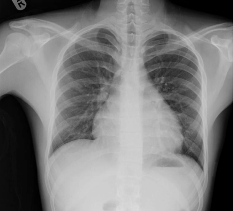
Figure 1: Chest x-ray showing globular cardiac silhouette(click to enlarge)
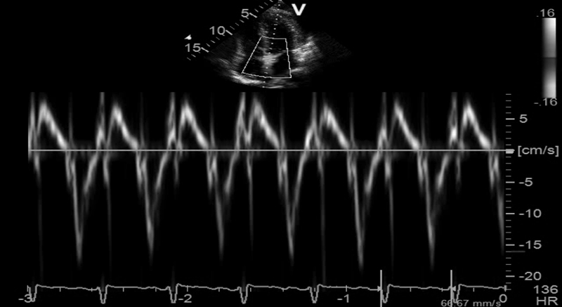
Figure 2: Tissue Doppler tracing high velocity myocardial excursions E’ @18 demonstrating pericardial constriction. Tissue Doppler is valuable in distinguishing constrictive from restrictive pericarditis showing high velocity excursions in the former and lower velocities in the latter; a cut off of 8 has been recommended by some authors(click to enlarge)
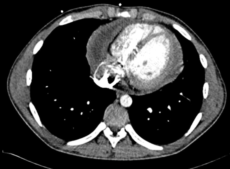
Figure 3: CT thorax showing a large pericardial effusion(click to enlarge)
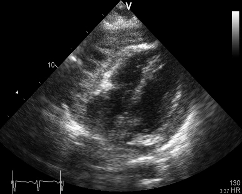
Figure 4: Subcostal view showing a thick exudative pericardial effusion. Thick fibrinous strands can be seen crossing the space and tethering the myocardium to the pericardium(click to enlarge)
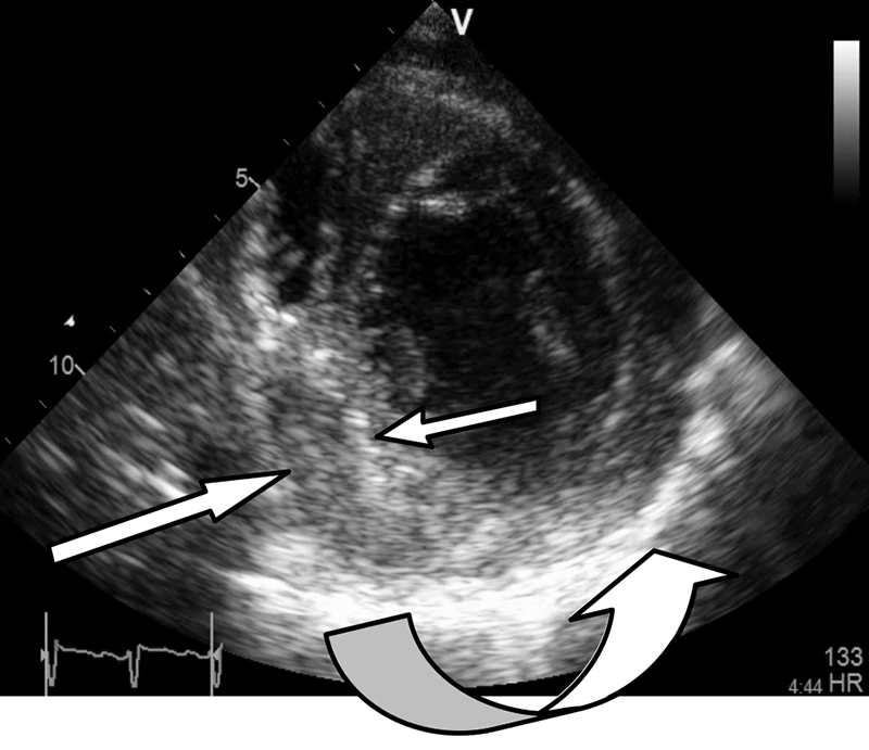
Figure 5: Short axis view showing a thickened hyper-echoic pericardium especially between 4 and 7 o’clock (curved arrow). Organised thick effusion indenting the infero-septal left ventricular wall between 6 and 9 o’clock (straight arrows)(click to enlarge)
Case report
A 20-year-old university student was referred to hospital by his GP with diminished effort tolerance of one month’s duration, an unexplained tachycardia and wide spread T-wave inversion on his ECG. On examination in the emergency department:
He was mildly pale, afebrile and anicteric
He was not orthopnoeic, dyspnoeic or tachypnoeic at rest
His lung fields were clear on auscultation
His JVP was not elevated
His blood pressure was 120/70
Cutaneous perfusion was normal and he did not have any pedal oedema
Abdominal examination was normal
No organomegaly or lymphadenopathy was evident
He admitted to having a slight cough and rather non specific left-sided chest pain but was otherwise well.
He was duly admitted to the medical assessment unit (MAU) on a Saturday afternoon for a work up. His management plan included admission into a monitored bay in the unit, a chest X-ray, FBC, ESR, CRP, U/E, D-dimers, a troponin cardiology consult and an outpatient echo.
Some 24 hours later another acute physician was approached by a junior doctor seeking permission to arrange a CT pulmonary angiogram and to commence anticoagulation on a 20-year-old with elevated D-dimers, a mild tachycardia and T-wave changes in the right ventricular leads – a reasonable request.
The consultant was struck by the fact that the patient’s heart rate was 110 when lying still in bed and that this increased to 130 when he attempted to sit up. His blood pressure was normal, he looked well, did not appear to have any risks for a pulmonary embolism or an obvious explanation for his tachycardia and markedly elevated D-dimers. His chest X-ray showed clear lung field but he had a rather globular ‘water bottle-shaped’ cardiac silhouette. The ECG revealed widespread T-wave inversion in all leads
The acute physician decided to perform a bedside echocardiogram to have an idea of RV function and pressures which might be abnormal in a significant PE and at LV function in case this was a cardiomyopathy.
Echo findings
Surprisingly the echocardiogram showed a large pericardial effusion containing thick viscid heterogenous fibrin rich exudate with evidence of septation loculation, and organisation, indicating that these changes were not acute. There was Doppler evidence of pericardial constriction; however, the absence of bi-atrial enlargement suggested that it might be fairly recent.
These findings are consistent with the widespread T-wave inversion on ECG, a feature of the later stages of pericarditis.
LV and RV function were moderately impaired with EF of 30-40% due to a combination of adhesion and tethering and constriction, hence the compensatory tachycardia to maintain cardiac output.
Diagnosis
A diagnosis of exudative pericarditis complicated by a large pericardial effusion and pericardial constriction was made. The patient had a departmental study on Monday which confirmed the portable echo findings.
An urgent cardiac CT and cardiac MRI were arranged, both of which confirmed the initial findings and revealed additional evidence of tethering pericardial constriction and even some pericardial calcification. His Mantoux test was reactive 14mm but he had prior exposure to BCG.
Findings were discussed at the cardiology echo conference and at the joint cardiothoracic multidisciplinary team meeting with a cardiac surgeon in attendance. The consensus was that needle paracentesis would be inadequate as the fluid was viscid loculated and at least partially organised, and that a tissue diagnosis to exclude tuberculosis would be helpful too. The possibility of a pericardiectomy was raised too.
The patient was transferred to a cardiothoracic centre for a video assisted thoracoscopic pericardial biopsy possibly as a prelude to a total or partial pericardiectomy, to prevent more severe pericardial constriction and its sequelae. Pericardial biopsy and analysis of the fluid confirmed a diagnosis of tuberculous pericarditis.
How did Echo change his management?
The diagnosis was made much sooner than it would have been if the patient had had an outpatient echo as planned initially. He appeared quite well and it was assumed that he had a viral pericarditis
An unnecessary CT pulmonary angiography (CTPA) was avoided
Therapeutic anticoagulation was not commenced. The echo had provide a valid alternative diagnosis to pulmonary embolism and therapeutic anticoagulation carries the risk of precipitating bleeding in to the pericardial sac which can cause cardiac tamponade
Anti-tuberculous therapy commenced sooner than if the patient had had to wait for an outpatient Echo cardiogram before the diagnosis was made, as suggested in his initial management plan.
Discussion
Pericardial constriction is a late complication of acute pericarditis, with grave implications for the patient if left undetected and untreated.
It results from the organisation/fibrosis of a protein rich inflammatory pericardial exudate that may encase the heart, impairing cardiac filling and emptying, eventually leading to symptoms and signs of right and or left heart failure.
The aetiology is usually viral but may also be due to tuberculosis (the latter condition has a mortality rate of up to 85% if untreated). Other inflammatory conditions such as connective tissue diseases, severe uraemia and cancer are culprits too. It is important to note that as this is an insidious process and patients may look and feel well due to prior to haemodynamic decompensation, inappropriate sinus tachycardia is a useful clue.
Echocardiography is probably the most efficient/effective way of making this diagnosis, especially from the perspective of the acute physician at the coalface.
Other techniques, such as cardiac MR and CT, can provide additional anatomical and functional detail. In general a multi-modality approach to imaging is advocated, especially if surgical intervention is planned.
Miniaturisation
Echo machines have become much smaller and modern laptop sized devices that often have capabilities that equal or exceed those of large departmental machines of a decade ago. These devices give the physician access to a treasure trove of information which is available immediately at the bedside any where in a hospital.
The portability of modern Echo machines is of particular value in acute medicine, a specialty in which choreographing flow is highly dependent on rapid access to diagnostic information about patients on the acute floor.
Early access to echo cardiographic information at the point of care about LV function, wall motion abnormalities, right ventricular dysfunction, the presence of tamponade or effusion, or significant aortic stenosis can be a significant game changer in critically ill patients, triggering transfer to intensive care, prompting thrombolysis, revascularisation or guiding fluid management.
As illustrated by this case, it can also speed up diagnosis and prompt appropriate management.
The ability to estimate LV function, identify RV dysfunction and severe valvular disease such as severe aortic stenosis in a breathless patient or in a patient with chest pain can significantly affect subsequent management.
Conclusion
Imaging technology needs to be used sooner on the acute floor. This has been recognised by forward-thinking medical assessment units and by colleagues in the specialty of emergency medicine for quite some time.
The author’s experience of the benefits of deploying echocardiography on the acute floor over the past five years prompts us to advocate training for acute physicians in basic echocardiography. Initially they should strive to be competent at performing focused studies to address specific questions such as the ones pertaining to LV function, RV function, pericardial effusion or tamponade.
Alternatively, and in order to address quality control issues, busy units should be provided with a dedicated or designated technician to perform echos on the hot floor on demand, in order to facilitate diagnoses, early discharge, risk stratification and to support ambulatory care.
In our experience the best solution is a synergistic hybrid that results from close co-operation between MAUs and cardiac diagnostics departments. In this model physicians and technicians perform echos on the acute floor and in the echo department, using a combination of portable and mainframe machines. Data from the portables re uploaded into the main departmental system at the end of the day for information governance, quality control teaching and future reference.
 Figure 1: Chest x-ray showing globular cardiac silhouette(click to enlarge)