Brain metastasis diagnosis from prostate carcinoma
Primary prostate cancer can cause pathologically confirmed brain metastasis. This article presents a recent case which demonstrates the phenomenon
Dr Ayman A Khalil, Specialist Registrar in Neurosurgery, Cork University Hospital, Cork, Dr Catherine Keohane, Consultant Neuropathologist, Cork University Hospital, Cork and Dr Charles Marks, Consultant in Neurosurgery, Cork University Hospital, Cork
Metastases from prostate cancer are common to the pelvic lymph nodes and the axial skeleton. Brain metastases from prostate primary are rare and most reported cases are from an autopsy series.1,2
Case report: brain metastasis presentation
A 60-year-old Irish man presented to the neurosurgery department with a one-month history of confusion and one-week history of progressive right-sided hemiparesis. He had a background history of poorly differentiated invasive adenocarcinoma of the prostate (Gleason score 7) diagnosed on prostatic needle biopsy, and had known widespread metastases in the bony skeleton for several years. He received palliative radiotherapy treatment locally to the bony skeleton metastases and was also receiving hormonal therapy (Prostap 3).
Investigations for the neurological symptoms included CT scan pre-contrast and post-contrast, which revealed a 6cm x 3cm complex mass lesion, in the left parietal-occipital area in the brain parenchyma with mass effect (see Figure 1).
Figure 1: CT brain scan showing 6cm x 3cm mass lesion (partially cystic) in the left parietal and occipital lobes (click to enlarge)
In addition, the isotope bone scan showed new bony metastases in the neck of femur bilaterally and in the thoracolumbar spine, and additional metastases in the bony rib cage and in the skull. The CXR demonstrated diffuse sclerotic metastases in the ribs. He underwent left occipital craniotomy and had subtotal removal of the intraparenchymal tumour, which at operation was partly cystic and contained clear brown fluid after decompression. It was not in continuity with skull metastases.
Histological examination of the resected tissue confirmed metastatic prostatic microglandular adenocarcinoma within the cerebral parenchyma, with strong positivity of tumour cells on immunohistochemistry for prostate-specific antigen (PSA [see Figure 2]) and showing areas of tumour necrosis.
Figure 2: A. Haematoxylin and eosin-stained section showing solid area of tumour composed of microglandular adenocarcinoma (10x);
B. Section immunostained with anti-prostatic specific antigen strong brown positivity in tumour cells and gland lumena (10x)(click to enlarge)
Post-operatively the patient was stable and, following multidisciplinary review, received palliative whole brain radiotherapy and also palliative radiotherapy to both hips. He died six months after brain surgery and did not have an autopsy.
Diagnosis and unusual detection
The diagnosis of intracranial metastases from adenocarcinoma of the prostate is relatively unusual. Most reported cases have come from autopsy series. In 1979, Catane et al found nine cases of intracranial metastases (1.1%) in 792 patients with adenocarcinoma of the prostate. In their autopsy series of 126 patients, Taylor et al found 14 patients (11%) with parenchymal intracerebral metastasis. A Japanese series revealed a prevalence of 2.1% of brain metastases.3
It seems likely that the low incidence may reflect both under-detection at autopsy and under-recognition in life due to the very late or pre-terminal occurrence of cerebral involvement, when investigation would be unlikely. Improved access to neuroimaging and improved survival from successful treatment of systemic complications may result in increased incidence.
The reported sites of intracranial prostate cancer metastasis are the leptomeninges (67%), the cerebrum (25%) and the cerebellum (8%).2
Usually, prostatic cancer patients with brain metastasis have widespread, advanced disease in other sites. In our patient, multiple metastases involving the brain and bone were detected four years after initial diagnosis of prostate cancer. In very few patients the brain is the only site of metastasis.5,6
It has been postulated that the mechanism of metastasis to the intracranial area may occur via direct access through the paravertebral venous plexus of Batson. This would avoid the bone and viscera. Or it may occur as part of a haematogenous spread in which initial metastatic foci in bone or lung might spread to involve the brain.7 In our case, while there was no direct connection to the skull metastases, haematogenous spread from a nearby bony metastatic focus may represent the mechanism of spread.
Neurological symptoms, which are secondary to the intracranial metastases as the first clinical manifestation of prostate cancer, are rare.6
This particular patient’s typical symptoms of an expanding destructive intracerebral lesion with mass effect led to the detection of the tumour.
Treatment options available for patients with brain metastasis from prostate cancer include radiation therapy or surgery. Data in the literature on survival after the diagnosis of brain metastases are limited due to the small number of antemortem cases.
In the past, patients with brain metastases from various primary sources had an overall poor prognosis, with one-year survival rate of 18% and an average survival of 7.6 months.5 However, patients with an isolated single metastasis have an improved quality of life and probable survival benefit if removed through surgery or radiosurgery.3 This patient survived six months following cranial surgery and removal of the metastasis to achieve symptom relief appears justified where the patient’s overall condition is sufficiently good to indicate a reasonable prospect of several months survival.
Conclusion
Intracerebral metastasis from prostate cancer is rare but likely to increase with improved patient survival from better therapeutic regimens. Surgery for solitary symptomatic cerebral parenchymal metastases can be successful in achieving palliative symptom control, and should be considered in a patient with prostate carcinoma who develops neurological symptoms.
References
Catane R, Kaufman J, West C et al. Brain metastasis from prostatic carcinoma. Cancer 1976; 38: 2583-2587
Lynes WL, Bostwick DG, Freiha FS, Stamey TA. Parenchymal brain metastases from adenocarcinoma of prostate. Urology 1986; 28: 280-287
Saitoh H, Hida M, Shimbo T et al. Metastatic patterns of prostate carcinoma. Cancer 1984; 54: 3078-3084
Gupta A, Baidas S, Cumberlin RK. Brain stem metastasis as the only site of spread in prostate carcinoma. A case report. Cancer 1994; 74: 2516-2519
Sutton MA, Watkins HL, Green LK, Kadmon D. Intracranial metastases as the first manifestation of prostate cancer. Urology 1996; 48: 789-793
Batson OV. The function of the vertebral veins and their role in the spread of metastases. Annals of Surgery 1940; 112: 138-149
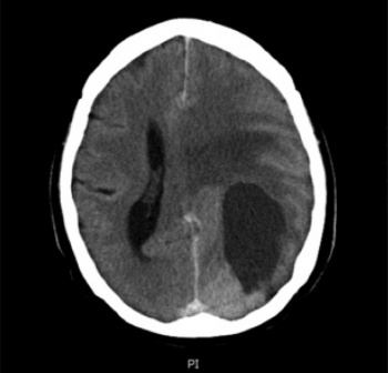 Figure 1: CT brain scan showing 6cm x 3cm mass lesion (partially cystic) in the left parietal and occipital lobes (click to enlarge)
