Carcinoma pharyngeal pouch discovered during surgery
A 78-year-old male patient presented with a six-month history of difficulty swallowing food and significant weight loss
Mr Sardar U Khan, Registrar, Department of ENT, Head and Neck Surgery, Midland Regional Hospital, Tullamore and Mr Leonard O'Keeffe, Consultant Otolaryngologist, Midland Regional Hospital, Tullamore
Pharyngeal pouch is a rare condition that occurs most commonly in older people. Typical symptoms include dysphagia, regurgitation of undigested food, chronic cough, aspiration and weight loss.
Carcinoma is a well recognised but rare complication of pharyngeal pouch (Zenker’s diverticulum). We report a case of carcinoma in a pharyngeal pouch diagnosed at endoscopy on the day of surgery. CT scan had shown symmetrical thyroid enlargement with kyphosis of the cervical spine, complicating surgical management. The treatment options available for this rare condition are reviewed and discussed.
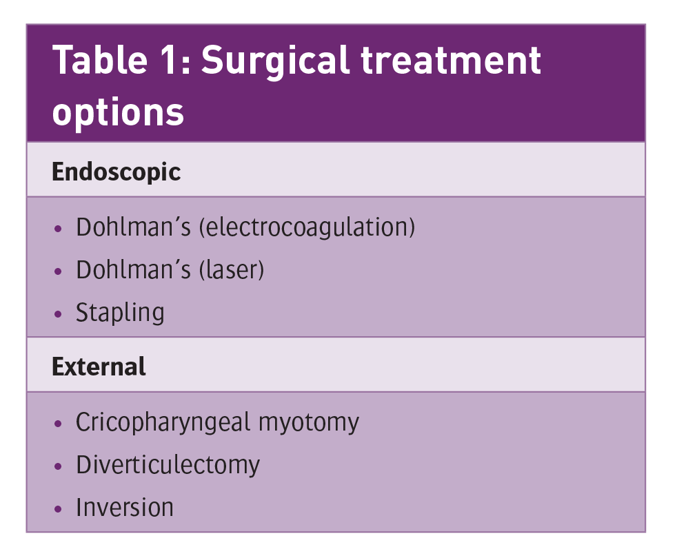
Table 1: Surgical treatment options(click to enlarge)
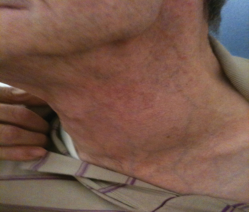
Figure 1: Swelling in the left lower neck(click to enlarge)
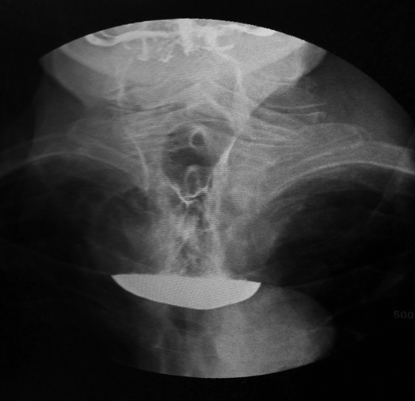
Figure 2: Barium study showing large pharyngeal pouch(click to enlarge)
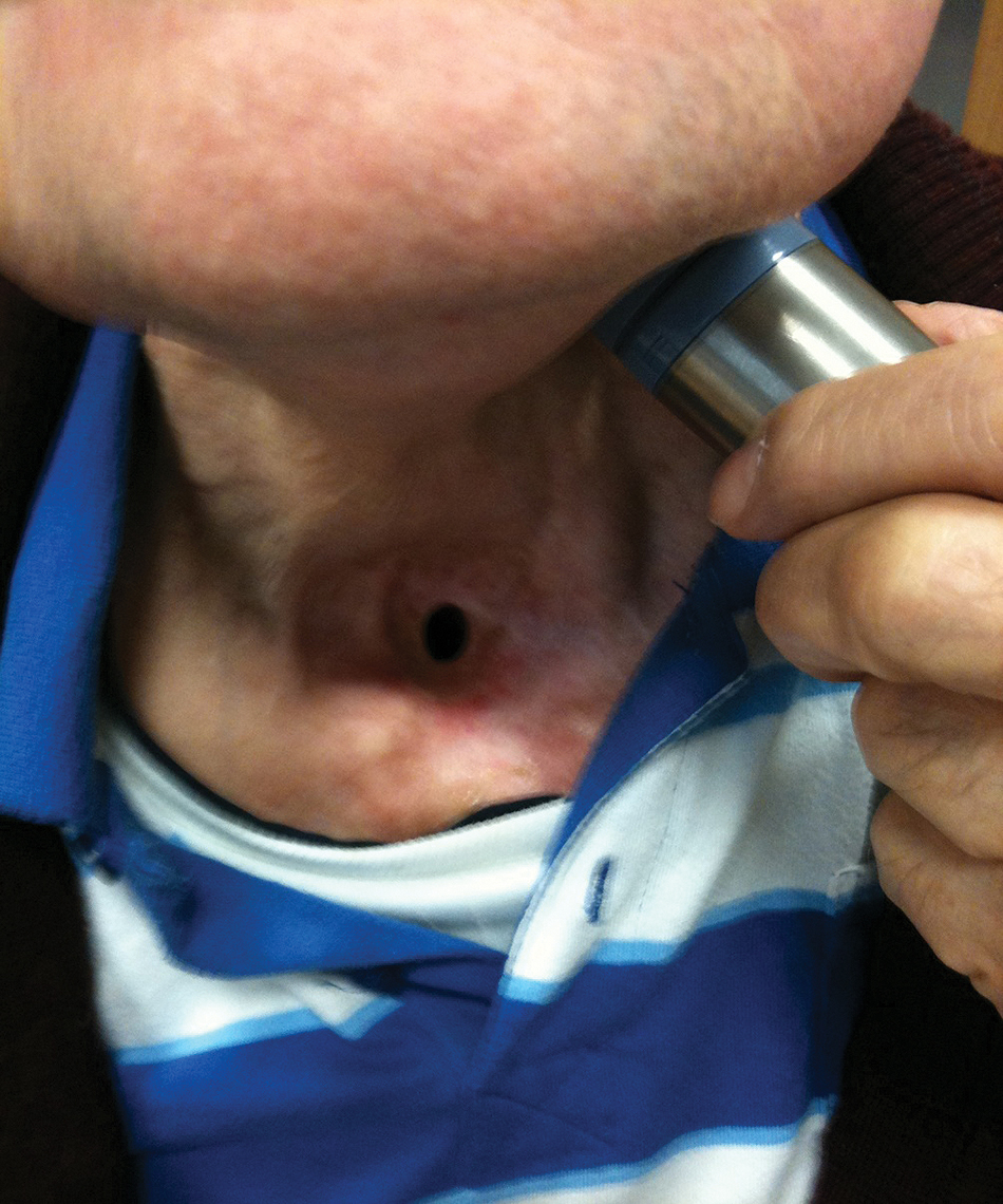
Figure 3: Patient using vibrator for speech(click to enlarge)
Introduction
Zenker’s diverticulum is an out-pouching of the pharyngeal mucosa through Killian’s triangle, an area of muscular weakness between the transverse fibres of cricopharyngeus and the oblique fibres of inferior constrictor muscle. The incidence is two per 100,000 of the population per year in the UK.1
The condition was first described by Ludlow in 1769. Later Zenker (1877) reviewed the presentation and pathology for this condition.2
The aetiology remains unknown but theories centre on a structural or physiological abnormality at the cricopharyngeal sphincter. Diagnosis is easily established on barium study. The treatment of choice for an established pharyngeal pouch is surgical and the approach may be external or endoscopic (see Table 1).
Case history
A 78-year-old male patient presented with a six-month history of difficulty swallowing solid food and significant weight loss. Recently the symptoms progressed to regurgitation of undigested food, with cough and a choking sensation. There was no pain or hoarseness.
Physical examination revealed swelling in the left lower neck (see Figure 1).
Flexiscope examination of the larynx and hypopharynx was normal. Contrast radiography demonstrated a large pharyngeal pouch (see Figure 2).
The patient was scheduled for excision of the pouch through an external approach. At endoscopy a large friable tumour was discovered at the level of cricopharyngeal sphincter. The lumens of the pouch and oesophagus could not be identified safely. Multiple biopsies were taken revealing invasive, moderately-differentiated, squamous cell carcinoma.
CT scan showed a 6x4cm mass occupying the left lower neck, displacing and compressing the oesophageal lumen to the right. Both lobes of the thyroid gland were enlarged. There was no adenopathy.
CT neck also demonstrated upper dorsal kyphosis with compensatory cervical lordosis, making surgical and anaesthetic management more difficult.
Definitive surgical management comprised total pharyngolaryngectomy, partial oesophagectomy, total thyroidectomy with bilateral neck dissection and jejunal free flap repair, followed by 40 Grays of postoperative radiotherapy.
Some three years after the procedure, the patient remains in remission with no dysphagia and using an artificial larynx (vibrator) for speech and communication (see Figure 3).
Discussion
Carcinoma is a rare complication of pharyngeal pouch (1%).3 A review in 1999 by Bradley et al, found 45 cases of pharyngeal pouch carcinoma reported in the English literature4 and reported two further cases. Since then one more case has been reported.5
The risk factors for the development of carcinoma in a longstanding pouch are: increasing age, chronic irritation and inflammation in the pouch.6 Neck compression and emptying of the pouch by the patient to assist swallowing may add to the irritation and contribute to the risk.
Malignant change in a pouch should be suspected when there has been sudden increase in the severity of symptoms, particularly worsening dysphagia, pain, haematemesis or sudden weight loss.3
This case presented with sudden increase in severity of dysphagia, more marked regurgitation of undigested food and weight loss.
Contrast studies rarely demonstrate a filling defect or loss of smooth contour of the interior of the pouch. In this case barium swallow showed a 4x6cm smooth-lined pouch with no suspicious radiological signs for malignancy.
Johnson and Curtin (1985) reviewed the radiology reports of 45 cases, and found that carcinoma was diagnosed in 14 (31%).7 Most cases are diagnosed at endoscopy, and oesophagoscopy should be performed in all cases, either as a separate procedure or as a prelude to definitive surgery.
In 13 of the reported cases, diagnosis has been established at endoscopy,3 as in our case. The histology is usually squamous cell carcinoma though variations such as basal cell or spindle cell carcinoma and carcinoma in situ have been reported.8,9
The usual treatment is one stage diverticulectomy followed by radiotherapy. When the neck of the diverticulum is involved, more extensive surgery in the form of laryngo-oesophagectomy is recommended.3
Endoscopic stapling is currently the procedure of choice for treating uncomplicated pharyngeal pouches. Anand reported the development of carcinoma in a recurrent pouch previously treated with endoscopic stapling.5
The patient was treated with excision of the pouch followed by radiotherapy. Therefore, excision rather than stapling should possibly be preferred in younger patients to avoid subsequent malignancy.
Conclusion
Carcinoma is a rare complication of pharyngeal pouch that tends not to be clinically or radiologically evident. Careful endoscopic examination of the diverticulum is diagnostic in most cases. The standard treatment is one stage diverticulectomy, followed by radiotherapy. When the neck of the diverticulum is involved, more extensive resection by pharyngo-laryngectomy is indicated.
References
Siddiq MA, Sood S, Strachan D. Pharyngeal pouch (Zenkers’s diverticulum). Postgrad Med J 2001; 77(910):506-11
Zenker FA, Von Ziemssen H. Krankheiten des oesophagus. Handbuch der speciellen pathologie and therapie. Vol 7; 1877: 1-87
Bowdler DA, Stell PM. Carcinoma arising in posterior pharyngeal pulsion diverticulum (Zenker’s diverticulum). Br J Surg 1987; 74:561-563
Bradley PJ, Kochaar A, Quraishi MS. Pharyngeal pouch carcinoma: real or imaginary risks? Ann Otol Rhinol Laryngol 1999; 108:1027-1023
Acharya A, Jennings S, Douglas S et al. Carcinoma arising in a pharyngeal pouch previously treated by endoscopic stapling. Laryngoscope 2006; 116(6):1043-5
Siddiq MA, Sood S. Current management in pharyngeal pouch surgery by UK otolaryngologist. Ann R Coll SurgEngl 2004; 86:247-252
Johnson JT, Curtin HD. Carcinoma associated with Zenker’s diverticulum. Annals of Otology, Rhinology and Laryngology 1985; 94:324-325
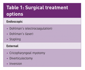 Table 1: Surgical treatment options(click to enlarge)