CARDIOLOGY AND VASCULAR

MENTAL HEALTH

DIABETES

ENDOCRINOLOGY

NEUROLOGY

Communicating hydrocephalus

The correct steps to take in the diagnosis and management of hydrocephalus

Dr Abuelmagd Abdalla, SHO in Medicine, Wexford General Hospital, Wexford and Dr Michael Conway, Consultant Cardiologist, St Luke's Hospital, Kilkenny

December 2, 2013

Article
Similar articles
  • Hydrocephalus can be defined broadly as a disturbance of formation, flow or absorption of cerebrospinal fluid (CSF) that leads to an increase in the volume occupied by this fluid in the central nervous system (CNS). This condition also could be termed a hydrodynamic disorder of CSF. Acute hydrocephalus occurs over days, subacute hydrocephalus occurs over weeks and chronic hydrocephalus occurs over months or years. 

    Conditions such as cerebral atrophy and focal destructive lesions also lead to an abnormal increase of CSF in CNS. In these situations, loss of cerebral tissue leaves a vacant space that is filled passively with CSF. Such conditions are not the result of a hydrodynamic disorder and therefore are not classified as hydrocephalus. An older misnomer used to describe these conditions was hydrocephalus ex vacuo.

    Case report 1

    A 63-year-old male who has a history of sero-positive rheumatoid arthritis (RA) on sulfapyrizine, was brought to the emergency department (ED) early in the morning unresponsive. He was found in bed at  home by his relative who noticed that his breathing was getting laboured and noisy. He was also found incontinent of urine with vomiting by his bed sheet. There was no seizure witnessed.

    He went to his bed normally the night before with no prior history of acute illnesses apart from his usual flares of rheumatic joint pains which are controlled with oral and parenteral non-steroidal anti-inflammatory drugs (NSAIDs) by his GP, although later a friend of the patient stated that he was texting him misspelled messages for the past two days, however, he wasn’t particularly confused or showing any behavioural change. His BP was 208/143 at scene by the ambulance crew. In the ED he was febrile and comatose (GCS 6/15) with no spontaneous limb movements. The initial CT brain was reported as  communicating hydrocephalus which was likely old with no other findings (see Figure 1).

    He was mechanically ventilated and remained in the intensive care unit (ICU) for the following 14 days. Investigations showed neutrophil leucocytosis and raised inflammatory markers in his blood. Meningoencephalitis was the working diagnosis from the beginning and he was covered with antimicrobial and antiviral from day one. His CSF analysis showed 35 WBC per/ul (95% lymphocytes), glucose of 3.5mmol (plasma 7.6mmol) and protein of 0.74g/L. Extensive microbiological analysis (including polymerase chain reaction [PCR]) failed to identify any organisms. The repeat CT brain three days later showed areas of low densities at right internal capsule and right temporo-parietal lobe, which are new since the first scan (see Figure 2) and which were suggestive of infarcts, and stroke was a possibility at that stage and was discussed with his family. The course of management, however, remained the same (completed full course of parenteral antibacterial and antiviral) and these areas totally disappeared in the later scans with no adequate explanation to their nature and implication.

    He subsequently improved with supportive measures: hydration and nutrition through nasogastric tube (NGT) feeding. He also retained his spontaneous breathing. He is currently maintaining adequate oral intake, having slurred speech and being incontinent of urine and faeces. He is fully dependent and requires full assistance on daily living activities due to residual weakness of all his limbs (grade 3/5 upper limbs, 2/5 lower limbs) and is awaiting a bed in the National Rehabilitation Centre.

    Figure 1. The initial CT brain at presentation showing the communicating hydrocephalus
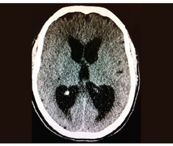
    Figure 1. The initial CT brain at presentation showing the communicating hydrocephalus(click to enlarge)

    Figure 2. The repeat CT showing the new areas of low attenuation (arrowed) which were thought to be infarctions but disappeared eventually
    Figure 2. The repeat CT showing the new areas of low attenuation (arrowed) which were thought to be infarctions but disappeared eventually(click to enlarge)

    Case report 2

    Our second case is a male of 59 years of age who was referred from the department of psychiatry with recent history (days) of deteriorating mobility and urinary incontinence. His past medical history includes long-standing intellectual disability,T2DM, hyperlipidaemia and depression. He is using oral hypoglycaemics, selective serotonin reuptake inhibitors (SSRIs), antidepressants, ACE inhibitors, hypnotics and olanzapine as long-term medications. 

    On further questioning, the psychiatry team and his family stated an ongoing history of being tearful, emotionally labile together with nihilistic ideation, reduced oral intake and lack of motivation of three weeks’ duration. No history of headache or vomiting. He was also independently mobile and continent of urine until a week before presentation. 

    His examination showed global rigidity but normal reflexes and sensation. No cerebellar or cranial nerve findings with intact upward gaze. Plantars were down-going. Normal peripheral nervous system and no autonomic involvement. His gait is apraxic with small steps and tendency to fall to both sides. No papilloedema encountered on fundoscopy.

    Investigations showed acute renal impairment likely secondary to his poor oral intake but no sepsis. His brain CT showed gross communicating hydrocephalus (see Figure 3). 

    There were no previous scans to compare and the finding is assumed to be new. Although part of his findings (rigidity, delusions and swinging mood) could be attributed to the use of second-generation (atypical) antipsychotics, this will not explain the hydrocephalus. The opening pressure of his CSF was 40cm/water and 50ml was removed as a therapeutic trial (after liaison with neurosurgery) to predict any possible benefit from the surgical CSF shunting. Unfortunately this hadn’t resulted in any significant improvement in his clinical status despite reduction in ventricular size on brain CT (see Figure 4). His CSF laboratory examination was essentially clear. 

    Other causes of hydrocephalus (eg. meningitis, subarachnoid haemorrhage [SAH], posterior fossa tumour) were looked for and excluded. He remained immobile except with assistance of 1-2, incontinent and intermittently agitated. He also needed full assistance for daily living activities apart from eating.

    Figure 3. CT brain showing gross communicating hydrocephalus
    Figure 3. CT brain showing gross communicating hydrocephalus(click to enlarge)

    Figure 4. The CT brain following removal of 50ml CSF. Note the reduction in ventricular size that did not correlate with clinical improvement
    Figure 4. The CT brain following removal of 50ml CSF. Note the reduction in ventricular size that did not correlate with clinical improvement(click to enlarge)

     Discussion

    Whilst the first case was likely post-meningitic and the second case was assumed to be idiopathic (one-third of total cases in the US are idiopathic), both were left with severe functional limitations. They are both totally dependent, incontinent and require full-time care. None of them will benefit from surgical interventions. They haven’t yet had seizure activity but epilepsy is an expected complication. Data on adult hydrocephalus are limited and it is always around treating the underlying cause whether found with consideration of surgical interventions (eg. shunting, third ventriculostomy) where appropriate. 

    While traditionally hydrocephalus used to be dealt with neurosurgically, today most of the cases are treated in other departments with referral only to those requiring interventions. The prognosis of adult-onset hydrocephalus is not usually positive, with the exception of those who receive timely diagnosis and intervention. Most are left with significant physical and mental disabilities requiring substantial rehabilitation. 

    © Medmedia Publications/Hospital Doctor of Ireland 2013