CANCER

CHILD HEALTH

Dual diagnosis of paraganglioma with acute liver abscess

An interesting case of dual diagnosis of an abdominal paraganglioma coincidental to an acute liver abscess in a 15-year-old boy

Dr Naveed Alam, Specialty Paediatric Registrar, Leicester Royal Infirmary, UK, Dr Mustaq Hussain, Consultant Paediatrician, North Cumbria University Hospital, UK, Dr Muhammad Rashid, Senior Paediatric Registrar, Portiuncula University Hospital, Galway, Dr Erik R Tjin A Ton, Consultant Radiologist, Portiuncula University Hospital, Galway, Dr Paula Cahill, Consultant Paediatrician, Portiuncula University Hospital, Galway and Dr Frances Neenan, Consultant Paediatrician, Portiuncula University Hospital, Galway

October 8, 2019

Article
Similar articles
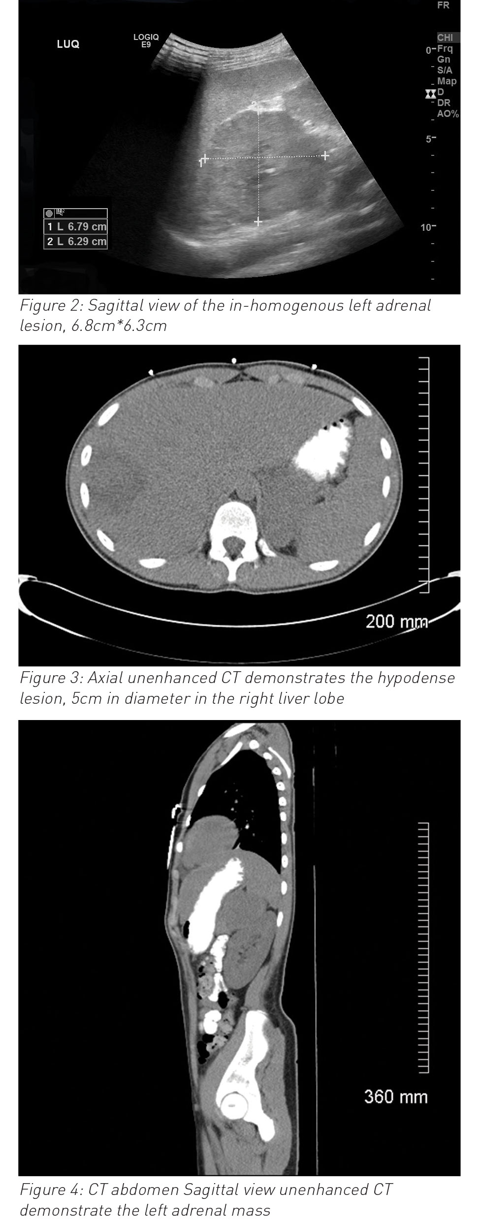
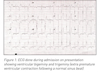
  • A 15-year-old boy presented with a short history of right upper quadrant abdominal pain, fever and right shoulder tip pain with high inflammatory markers. Within 24 hours he developed ventricular bigeminy and trigeminy. An abdominal ultrasound showed a liver abscess and unexpected findings of left side suprarenal mass. Subsequent MRI abdomen, MIBG scan and histopathology confirmed the diagnosis of an abdominal paraganglioma coincidental to the acute presentation of a liver abscess, likely from preceding trauma.

    On presentation to the emergency department, this previously healthy adolescent had a one-week history of being unwell, vomiting, arthralgia, myalgia and poor appetite which was preceded by coryzal illness for few days. The following day he started complaining of abdominal pain and right-sided shoulder tip pain and low-grade pyrexia. He had a history of a significant fall from a petrol go-kart while racing two weeks earlier with bruising to his back and abdomen.

    On clinical examination, he was unwell, pale, tachycardia (H.R: 130/min), pain score: 8/10, positive Murphy sign. Blood investigation showed high inflammatory markers with white blood cells of 36.4 x 10⁹/L and C-reactive protein of 252mg/L) normal liver functions test and serum amylase. The working diagnosis of acute cholecystitis established and was commenced in intravenous antibiotics (initially on co-amoxiclav and then switched to cefotaxime) and further radiological investigations were requested.

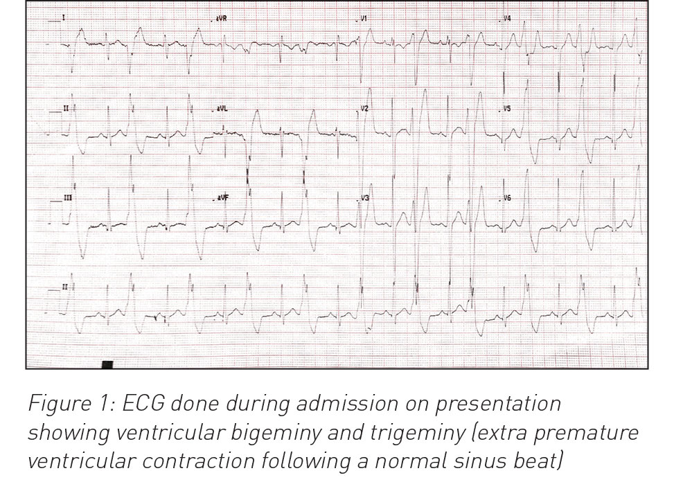
    During his hospital stay it was noted that he had an episode of high blood pressure (140/90mmHg) and developed ventricular bigeminy and trigeminy (see ECG in Figure 1). Chest x-ray and echocardiography were normal. Ultrasound and MRI abdomen reported left side suprarenal mass (8cm) and associated liver abscess (7cm x 7cm x 7cm). Urine catecholamines were normal. Liver abscess (abscess grew Streptococcus intermedia) was treated with drainage and with six weeks antibiotics. Paraganglioma was surgically resected successfully three months after initial diagnosis and full evaluation (genetic testing and exclusion of MEN syndrome and von-Hipple-Lindau syndrome). Ventricular bigeminy resolved completely and final diagnosis of non-secreting pheochromocytoma and coincidental liver abscess (likely due to trauma in the past). He was discharged home after full recovery. He is being followed up with regular abdominal imaging; to date, no concerns have been reported.

     (click to enlarge)

     (click to enlarge)

    Discussion

    Pheochromocytoma/paragangliomas (PHEO/PGL) are quite rare in paediatric patients with an incidence of ≤ 0.3 cases per million per year. Some 10-20% of cases are diagnosed during childhood, with the average age of presentation being 11 years, with slight male predominance.1

    Roughly 22% of PHEO/PGL are extra-adrenal. Paragangliomas are hugely vascular lesions that originate from neural crest cells, which are catecholamine secreting cells.2 Most of the time the paraganglioma is right-sided adrenal in location, but is also rarely found in liver, pancreas and retroperitoneal.2,3,4

    The majority are reported as benign tumours, however roughly 12% of  tumours are malignant which can metastasise to surrounding tissues.1

    Useful tests include CT or MRI of abdomen and pelvis, imaging of the neck and chest, urine catecholamine and USG. However, an MIBG scan is a highly specific test for diagnosing catecholamine-secreting tumour.1

    The prognosis of a paraganglioma is excellent after a complete resection.1

    Prior to surgery 1-alpha adrenergic blockade is used,1 in some cases phenoxybenzamine (non-competitive alpha-blocker) is also used.1

    Follow up for at least three years is necessary as PHEO/PGL can have unpredictable behaviour.1,2 These tumours can also metastasise later after surgery. Some patients may be at risk for the development of meta-chronous tumours, so long term follow-up is recommended.1

    Conclusion

    This case is an excellent example of interdisciplinary team work that resulted in improved patient outcome. Bringing such an unusual presentation to the attention of general paediatricians may prompt them to consider a  dual diagnosis or co-occurring pathology to solve a clinical dilemma. 

    References

    1. Waguespack SG, Rich T, Grubbs E, Ying AK, Perrier ND, Ayala-Ramirez M, Jimenez C. A current review of the etiology, diagnosis, and treatment of pediatric pheochromocytoma and paraganglioma. J Clin Endocrinol Metab 2010 May; 95(5): 2023-2037
    2. Khan MR, Raza R, Jabbar A, Ahmed A. Primary non-functioning paraganglioma of liver. J Pak Med Assoc 2011 Aug;61(8):814-816.
    3. Furcea L, Mois E, Al Hajjar N, Seicean A, Badea R, Graur F. Pancreatic gangliocytic paraganglioma - CEUS appearance. Journal of Gastrointestinal and Liver Diseases 2017; 26(4): 336
    4. Pandurengan K, Natarajan R, Marudavanan R, Sowmya T, Raja chidambaram K. An unusual retroperitoneal mass - non functioning retroperitoneal paraganglioma. Internet Journal of Endocrinology 2011;6(2). http://ispub.com
    5. Melicow MM. One-hundred cases of pheochromocytoma (107 tumors) at the Columbia-Presbyterian Medical Center, 1926-1976: a clinicopathological analysis. Cancer 1977 Nov;40(5):1987-2004
    © Medmedia Publications/Hospital Doctor of Ireland 2019