GASTROENTEROLOGY

Haemobilia: symptoms, diagnosis and treatment

Haemobilia, a rare cause of upper gastrointestinal (GI) bleeding following liver biopsy

Dr Ammar Shahin, Gastroenterology Registrar, Sligo General Hospital, Sligo and Dr Kevin Walsh, Consultant Gastroenterologist, Sligo General Hospital, Sligo

February 10, 2014

Article
Similar articles
  • A male in his early 70s who has a background history of hypertension, renal cell carcinoma (RCC) post-left nephrectomy five years ago, was referred to the gastroenterology clinic for evaluation of mildly persistently elevated liver enzymes.

    Case report

    Initial work-up, including hepatitis profile and autoimmune screen, was negative. His liver functions including bilirubin, albumin and international normalised ratio (INR) were normal. Subsequently, he was scheduled for percutaneous liver biopsy which was done under ultrasound guidance. Two days after the biopsy he developed right upper quadrant abdominal pain, jaundice and black stool and so was admitted to the hospital. His haemoglobin dropped to 7.5 so he was transfused with three units of red blood cells. Abdominal ultrasound showed blood within the biliary system. He was managed conservatively and the haemoglobin remained stable, the abdominal pain resolved and he was discharged home. Two days later he was readmitted with worsening abdominal pain.

    Upper GI endoscopy done during this admission showed blood within the distal stomach, and more fresh blood within D1 and D2 with blood coming from the ampulla of Vater. This confirmed the diagnosis of haemobilia. The liver biopsy was reported as mild inflammation and fibrosis without significant cirrhosis.

    Figure 1. Blood coming from ampulla of Vater at upper GI endoscopy
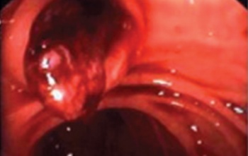
    Figure 1. Blood coming from ampulla of Vater at upper GI endoscopy (click to enlarge)

    Figure 2. Blood within the biliary ducts
    Figure 2. Blood within the biliary ducts (click to enlarge)

    As a result the patient was transferred to the liver unit at St Vincent’s University Hospital, Dublin, where he underwent hepatic artery embolisation and insertion of coils. Unfortunately the bleeding continued and so the angiogram and the embolisation were repeated with more coils inserted; the bleeding stopped and he was discharged home. Following the second angiogram his haemoglobin stabilised at 11.5 without any need for further intervention. 

    Haemobilia, bleeding that occurs from the hepatobiliary tract, is an extremely rare complication of liver biopsy that causes acute upper GI bleeding. Its incidence is 0.058-0.2%.1 It should be considered in patients who have a recent history of hepatic or biliary tract injury. 

    This includes trauma to the area and percutaneous (or transjugular) liver biopsies, and percutaneous transhepatic cholangiograms or biopsies.2 Haemobilia can also occur from gallstones, cholecystitis, hepatobiliary tumours, hepatic abscesses and aneurysms. The classic triad of haemobilia is biliary colic, obstructive jaundice and GI bleeding presents in about one-third of patients. Obstructive jaundice may be associated with biliary sepsis and cholangitis. The diagnosis of haemobilia can be made by ultrasound, magnetic resonance cholangiopancreatography (MRCP); the diagnosis by upper GI endoscopy may be difficult. A side-viewing duodenoscope is very helpful to look at the ampulla directly by MRCP or to perform endoscopic retrograde cholangiopancreatography (ERCP), tagged red blood cell scan or angiography can be done to localise the source of bleeding.3 Treatment is directed at the primary cause of bleeding. 

    The patient may respond to conservative management with blood transfusion and monitoring the blood count as spontaneous recovery is usual. Although occasionally treatment can be carried out endoscopically by injection of epinephrine at the ampulla, it usually needs to be done angiographically (such as by arterial embolisation) or surgically if embolisation failed, which includes arterial ligation. Surgical intervention is also indicated if there is biliary sepsis or if drainage failed.4

    References

    1. Dooley JS, Lok ASF, Burrougjs AK, Heathcote J. Sherlock’s diseases of the liver and the biliary system, 12th ed, p 285
    2. Hauser SC, Pardi DS, Poterucha JJ. Mayo Clinic Gastroenterology and Hepatology Board Review, 3rd ed, p 339
    3. Greenberger NJ, Blumberg RS, Burakoff R. Current diagnosis and treatment gastroenterology, hepatology and endoscopy, 2009, p 338
    4. Bloechle C, Izbicki JR, Rashed MY et al. Haemobilia: presentation, diagnosis and management . Am. J. Gastroenterology 1994; 89: 1537–1540w
    © Medmedia Publications/Hospital Doctor of Ireland 2014