WOMEN’S HEALTH

Management of mastocytosis throughout pregnancy

A clear management plan for the antenatal period, labour and postnatal period should be devised for the patient

Dr Irum Farooq, SHO in Obstetrics and Gynaecology, the Coombe Women and Infants University Hospital, Dublin and Dr Karen Flood, Consultant in Obstetrics and Gynaecology, Rotunda Hospital, Dublin

June 24, 2016

Article
Similar articles
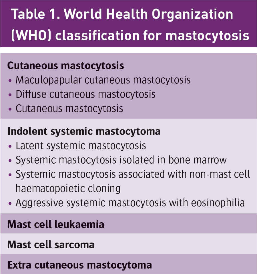
  • Mastocytosis is a rare disease, characterised by pathologic increase in mast cells in cutaneous tissue and extra cutaneous organs such as the bone marrow, liver, spleen and lymph nodes. It comprises many different clinical presentations varying from indolent cutaneous forms to malignant and systemic conditions. Episodes of life-threatening anaphylaxis are a recognised feature of the disease. This case report highlights the management of cutaneous and systemic mastocytosis in pregnancy.

    A 28-year-old primigravida booked for antenatal care in the hospital at 28 weeks gestation. She had no significant family history. She had recently been diagnosed with cutaneous mastocytosis. Mastocytosis work up was performed because she collapsed twice while hiking in mountains seven years ago. She had no apparent systemic reaction, however she was hospitalised and required epinephrine following the first event. There were no further events for seven years. 

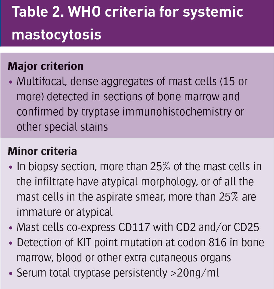
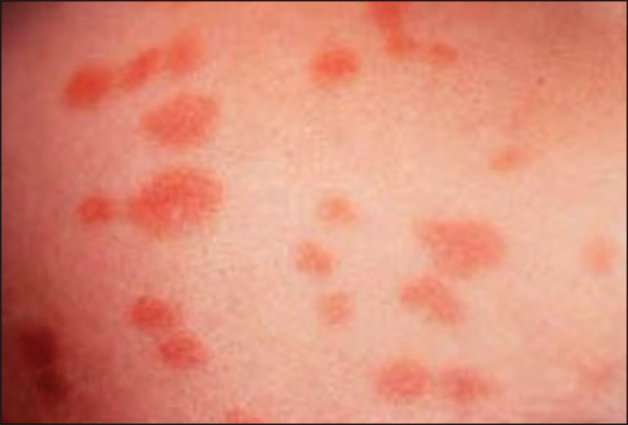
    She gave a history of a cutaneous rash for at least 10 years, small pinkish light brown papules which become erythematous with irritation or sun exposure. She also had a history of intermittent flushing and urticaria. Her diagnosis was based on pathological examination of a skin biopsy which demonstrated a mast cell infiltrate in the upper dermis consistent with urticaria pigmentosa. She also had elevated tryptase level, a marker of mast cell degranulation released in parallel with histamine. She had negative CKIT D816V mutation, which was reassuring as far as risk to future offspring was concerned. 

    Of note, most forms of mastocytosis are caused by a mutation of the KIT gene on the 4q12 chromosome – a mutation which increases cellular reproduction. Mastocytosis can be associated with allergic and anaphylactic reaction; consequently she has been prescribed an EpiPen in case of an anaphylaxis by her physicians. 

    When the woman booked for antenatal care she was referred to a high risk antenatal clinic, which included regular foetal biometry assessment, and attendance at specialised anaesthetic and haematology clinics. Her case was discussed at our monthly multidisciplinary meeting (also attended by lead hospital pharmacist) and at 36 weeks gestation a final plan of care was devised and circulated to relevant personnel. The team reviewed all available data and agreed that when she presented in labour, she should receive 100mg IV hydrocortisone (with consideration of six hourly hydrocortisone also) and should be given PO antihistamine, eg. chlorphenamine 10mg, IV. A list of histamine releasing drugs to be avoided included morphine, codeine, aspirin, suxamethonium, atracurium and rocuronium. Non-steroidal drugs to be avoided included diclofenac sodium (Difene), ibuprofen and mefenamic acid (Ponstan). Drugs which were considered safe for use included propofol, ketamine, fentanyl, sevoflurane and paracetamol. Early epidural was recommended in order to avoid a general anaesthetic. Consultant involvement at all times was also recommended. It was decided to involve a consultant anaesthetist if the woman required a cesarean section and to be transferred to high dependency unit postpartum. The plan for post-operative pain was for an epidural infusion for 24 hours and fentanyl via patient controlled analgesia to help control her pain. In the case of postpartum haemorrhage oxytocin was to be used. If there was any evidence of infection clindamycin and ciprofloxacin were considered safe to be used, however vancomycin was to be avoided. There was also a consideration for the use of high sensitivity gloves. Most importantly, it was agreed that adrenaline should be readily available. If she became hypotensive following epidural administration epinephrine should be administered. 

    Ultimately spontaneous labour occurred at 40+4 weeks and she used entonox and received an early epidural as planned. A female baby weighing 4.18kg was delivered by spontaneous vaginal delivery with a second degree tear occurring, which was sutured. She was transferred to the high dependency unit postpartum. Neither local anaesthetic agents nor antibiotic agents caused any reaction. The postpartum period was uneventful.

     (click to enlarge)

     (click to enlarge)

    Discussion

    The prevalence of mastocytosis is reported to be one in 1,000-8,000. Mastocytosis occurs in all races, with no sex predilection. Peak incidence is in infancy and early childhood, with a second peak in middle age. In some cases, the genetic disorder is inherited but in most cases it is sporadic and with no family history of mastocytosis. Symptoms occur when triggers such as drugs, exercise, stress, anxiety and temperature extremes cause mast cell degranulation and release of histamine, prostaglandins, leukotrienes, tryptase and other chemical mediators. Anaphylactic symptoms in adults with mastocytosis are primarily cardiovascular in nature with dizziness, presyncope, tachycardia and hypotension or shock. Symptoms can be observed in the skin (urticaria, angioedema, flushing), mucosal tissue (swelling of the tongue, uvula and intestinal mucosa resulting in dysphagia, abdominal cramps, vomiting and diarrhoea), respiratory tract (dyspnoea, wheezing, stridor, reduced peak flow and hypoxaemia), cardiovascular system (drop in blood pressure, syncope), and psychological symptoms such as irritability and difficulty in concentration may also be associated with the disorder.

    Cutaneous mastocytosis constitutes 90% of cases of mastocytosis not associated with a haematologic disorder. Urticaria pigmentosa is the most common variety, occurring in about two-thirds of patients with cutaneous mastocytosis. Tan to red-brown macules typical of urticaria pigmentosa initially emerge on the trunk and rapidly spread symmetrically and centripetally. The palms, soles, face and scalp are usually spared. The lesions may remain small and freckle-like or may evolve into papules, nodules or plaques (see Figure 1). Mucous membranes may also be involved. Exposure to physical stimuli such as heat, cold or pressure is typically followed by the onset of localised urticaria (Darier’s sign). Scarring is unusual unless super infection occurs.

    Figure 1. Cutaneous mastocytosis furticaria pigmentosal
    Figure 1. Cutaneous mastocytosis furticaria pigmentosal(click to enlarge)

    Diagnosis is often difficult and delayed due to variety of presenting symptoms. It can be diagnosed by taking a detailed history and physical examination but usually a skin biopsy is a necessary confirmatory test. 

    Patients with symptomatic mastocytosis should identify and avoid triggering factors. Extremes of temperature, alcohol and certain drugs should be avoided completely or used with caution. Pharmacologic treatment of mastocytosis involves stabilising mast cell membranes to decrease the severity of the attacks while blocking the action of inflammatory mediators. Histamine H1 antagonists are used to treat pruritus, flushing, bullae and urticaria. Histamine H2 blockers can be used to decrease the gastric hyperacidity. Patients with mastocytosis should also wear a medical alert bracelet and carry an adrenaline-filled syringe. The emergency treatment of shock in association with mastocytosis is the same as that of anaphylaxis. Fluids, adrenaline, antihistamines and vasopressor agents are often required.

    Conclusion

    Pregnant women with mastocytosis should be treated symptomatically and educated to avoid factors that may exacerbate symptoms of disease. Patients should obtain oral and written instruction and know proper techniques for self-administration of epinephrine in case of anaphylaxis. A clear management plan for the antenatal period, labour and postnatal period should be devised for patient. A multidisciplinary approach is essential including early involvement of senior obstetrician, haematologist, pharmacist, anaesthetist and microbiologist. As a preventive measure, resuscitation equipment should be available during the labour, delivery and postpartum period to treat unanticipated hypotension and shock.

    References
    1. Alto WA, Clarcq L. Cutaneous and systemic manifestations of mastocytosis. Am Fam Physician 1999; Jun 1; 59(11); 3047-3054
    2. Hogan DJ. Mastocytosis. Nova South-Eastern University College of Osteopathic Medicine; Investigator, Florida Research Centre. Feb 2015 http://emedicine.medscape.com/article/1057932-clinical
    3. Maluf LC, Barros JA, Machado Filho CD. Mastocytosis. An Bras Dermatol 2009; 84(3): 213-25
    4. Brockow K, Jofer C, Behrendt H, Ring J. Anaphylaxis in patients with mastocytosis: a study on history, clinical features and risk factors in 120 patients. Department of Dermatology and Allergy Biederstein, Technical University Munich; Division of Environmental Dermatology and Allergy GSF/ TUM, Munich, Germany, September 19, 2007
    5. Worobec AS, Akin C, Scott LM, Metcalfe DD. Mastocytosis complicating pregnancy. Obstet Gynecol 2000 Mar; 95(3): 391-395
    6. Hennessy B, Giles F, Cortes J et al. Management of patients with systemic mastocytosis: Review of MD Anderson Cancer Center Experience. American Journal of Hematology 2004; 77:209-214
    7. Müller UR, Haeberli G. The problem of anaphylaxis and mastocytosis. Current Allergy and Asthma Reports 2009; 9:64-70
    © Medmedia Publications/Hospital Doctor of Ireland 2016