A case of life-threatening menstrual staphylococcal toxic shock syndrome
Dr Ciarán McDonald, Senior House Officer (Infectious Diseases), Cork University Hospital, Cork, Ms Julia Marr, Final-Year Medical Student, University College Cork, Cork, Dr Cathal Ó Broin, Infectious Diseases Specialist Registrar, Cork University Hospital, Cork and Prof Mary Horgan, Consultant in Infectious Diseases, Cork University Hospital, Cork
A 15-year-old girl with GP referral presented acutely unwell for less than 24 hours to the emergency department (ED) and was admitted under the paediatrics service. She had awoken that morning with two episodes of vomiting with a further 10 episodes noted the previous day. There was no haematemesis and the vomitus was described as bilious-like. She was not tolerating any oral fluids and had three episodes of profuse foul-smelling, watery diarrhoea the previous day. She was pyrexic with rigors overnight and exhibited a generalised macular blanching rash. She complained of gradual onset headache and generalised leg pains. There was no photophobia or overt nuchal rigidity. On examination she was able to weight bear. On attending the GP that morning, she was hypotensive at 60/40mmHg and her capillary refill time was six seconds with a serum glucose level of 1.8mmol. She received a stat dose of glucagon along with 2ml/kg dextrose 10% IV. Pre-hospital antibiotics administered included 1g ceftriaxone IV, 750mg cefuroxime IV and 1,200mg benzylpenicillin intramuscularly. Overall, she received two boluses of IV NaCl 0.9% (20ml/kg) in the GP surgery prior to hospital admission.
Her background medical history included an injury to her right eye resulting from trauma while playing with a bat several months earlier. This resulted in a chronically dilated pupil and lens dislocation of the affected eye. This was under conservative review with ophthalmology. Her regular medications included brinzolamide timolol (Azarga) and dexamethasone (Maxidex) eye drops, both topically applied to her right eye.
The patient was delivered term +13 days by vacuum delivery with no complications. All vaccinations were up-to-date and the patient had reached all development milestones appropriately. Of note, she had childhood asthma, which had been quiescent for the past couple of years. She lives at home with her mother and two siblings and attends secondary school. She has no notable sick contacts. She is a non-smoker, non-drinker. There was no recent foreign travel.
On examination in the ED, her vitals were as follows: respiratory rate (RR) 24, oxygen saturation 97% on 4L oxygen, heart rate (HR) 125bpm regular, BP 111/68, temperature 38.2˚C. Her Glasgow coma scale (GCS) was 15/15 and the patient was alert and interacting appropriately. Cardiovascular exam revealed audible S1 S2 with no added heart sounds or murmurs. Her peripheries were cool but improving with IV fluid resuscitation. Her respiratory exam revealed audible bibasal breath sounds with no crackles, wheeze or stridor noted. Abdomen was soft and non-distended with mild generalised tenderness most notable in right upper quadrant (RUQ). Bowel sounds were present. An extensive blanching erythematous rash was noted over the chest, abdomen and medial aspect of thighs. Perioral examination showed no overt erythema or swelling of pharynx, visible exudate or stigmata of measles. Cervical lymph nodes were normal bilaterally. Neurological examination was normal for tone, power, reflexes and sensation throughout.
Initial blood work revealed a prominent leukocytosis (WCC 18.2, neutrophils 17.92, lymphocytes 0.15). Hb was stable at 14.7g/dl, and platelets were 142. CRP was markedly elevated at 240 with a lactate of 4.9mmol/L and a pH of 7.28. She had an acute kidney injury with urea 16.4μmol/L and creatinine 435μmol/L. She was coagulopathic with an INR of 2 and her PT 21.5 sec. A urinary catheter was inserted for fluid balance management. Initial urinalysis showed ++ protein and a trace of leukocytes. Her beta hCG was negative. Pelvic vaginal (PV) examination under aseptic technique revealed no foreign bodies with a swab taken in light of a yellow liquid discharge noted on close inspection. Her chest x-ray showed no acute findings with any visible effusions. A portable abdominal ultrasound was performed which showed bilateral medical renal disease with no acute obstructions. Allied to this was a normal appearing liver with no abscesses or cysts. Sludge was seen in her gallbladder but no cholecystitis. The appendix was not visualised and a CT TAP was arranged. The CT TAP showed no major acute focus of infection. Small sub centimetre ill-defined ground glass nodules were seen in both lungs. Her appendix was prominent but did not show any acute signs of appendicitis.
The patient remained in persistent hypotensive shock with BP 78/43mmHg despite 20ml/kg IV fluid boluses over the initial three hours in ED (a total of >3L over a six hour period). She was reviewed by the renal service with a view to commencing hemofiltration. In addition to her pre-hospital antibiotics, 180mg gentamicin (3mg/kg) and 900mg vancomycin were administered. Her peripheries became more mottled with capillary refill time of three seconds centrally. A decision to intubate and artificially ventilate was made as the patient remained in persistent septic shock and she was transferred to the ICU.
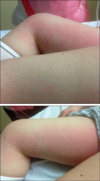
Her lactate continued to improve from 4.9 to 1.8 mmol/L with the IV fluids. However, she remained profoundly in a metabolically acidotic state with a pH of 7.22 and HCO3 of 15.6. In ICU the patient stabilised with her BP rising to a steady level of 150/70mmHg with phenylephrine. MAP was maintained >65mmHg with vasopressors and BGL >4mmol/l with 5% dextrose at 50ml/hr. Her lactate on serial ABG sampling showed good resolution to 2.6 after an initial 16 hours of treatment. The infectious diseases team was consulted while the patient was in ICU. The likely diagnosis was felt to be a toxic shock syndrome secondary to either Staphylococcus or Streptococcus and acyclovir was accordingly discontinued. The rash, which had previously been more extensive, was now more localised to the groin streaking upwards medially to the inguinal region bilaterally and had a sunburn appearance (see Figures 1 and 2).
Figures 1 and 2. The rash, which had previously been more extensive, became more localised to the groin streaking upwards medially to the inguinal region bilaterally and had a sunburn like appearance(click to enlarge)
A high vaginal swab (HVS) was performed which was sent for virology exotoxin testing for TSST-1 and TSST-2 along with culture and sensitivities. ASOT, EBV, CMV, complement 3 and 4 along with urinary pneumococcal antigen were added. Results were still pending from a full autoimmune screen. Blood cultures had been taken from admission but repeat cultures were taken as the patient continued to spike temperatures to 38˚C cyclically.
The patient made a good recovery over the coming days with extubation and weaning down of vasopressors occurring 48 hours after initial admission to ICU. Her renal function improved without any haemofiltration from a creatinine level of 435μmol/L to 108μmol/L. Her lactate had also resolved to normal levels with the coagulopathy correcting itself over time. Her LFTs remained slightly deranged; this was attributed to her antibiotics. The patient was transferred to the paediatrics ward 72 hours after initial presentation to hospital.
Cultures from her vaginal swabs had returned positive for MSSA and her antibiotics were rationalised to clindamycin 450mg PO BD (7mg/kg) and flucloxacillin 2g IV QDS. The patient encountered a drug-induced erythematous rash on the ward after her second dose of flucloxacillin and this was discontinued with vancomycin added back in.
The patient continued her quick recovery with CRP returning to 16 five days after admission. There was some desquamation of skin that had previously been affected by the erythematous rash and some small amount of hair loss was noted over affected areas. The patient remained apyrexial throughout her admission. She was discharged after a normal ECHO nine days after admission. A social worker had engaged with both the patient and parent regarding personal hygiene at the time of menstruation.
Meanwhile, the HVS sent while in ICU had returned positive for toxic shock syndrome toxin 1 and 2. This confirmed the diagnosis of toxic shock syndrome secondary to methicillin sensitive Staphylococcus aureus, likely from foreign body use around the time of menstruation. She was discharged home on clindamycin 450mg PO BD for a total course of 14 days. Follow up in clinic after this had revealed a complete and full recovery with normalisation of all blood work.
Background
S. aureus is a gram-positive bacteria that colonises the skin and mucous membranes in the anterior nares, vagina and rectum in 30-50% of the healthy population.1 It is an opportunistic pathogen with an arsenal of virulence factors that enables it to cause a broad range of disorders. It is associated with inflammatory diseases such as cellulitis, impetigo, abscesses, pneumonia, infective endocarditis, septic arthritis, osteomyelitis and septicaemia, as well as toxin-mediated diseases such as food poisoning, scalded skin syndrome and toxic shock syndrome. S. aureus is a leading cause of disease in both hospital and community settings.2 Emergence of antimicrobial-resistant strains such as methicillin resistant S. aureus (MRSA) demonstrates the bacteria’s ability to evolve. Systemic MRSA infections are difficult to treat and are associated with a high mortality rate, making it a health concern of increasing importance.2
Staphylococcal toxic shock syndrome (SaTSS) is a fulminant infection due to S. aureus. The majority of cases of SaTSS are due to methicillin sensitive S. aureus (MSSA) but incidence of MRSA SaTSS is increasing due to the increasing prevalence of MRSA. The initial clinical presentation of SaTSS is non-specific with high fever, sunburn-like rash, hypotension, myalgia, confusion and gastrointestinal symptoms, followed by desquamation of the skin one to two weeks after onset of disease. There is no single diagnostic test for SaTSS and diagnosis of SaTSS is therefore difficult to make due to its vague clinical picture.
SaTSS was first documented in 1978. The incidence increased drastically in the following years, during which time, the majority of presentations were in young women. SaTSS was eventually found to be associated with the use of high absorbency tampons and changing tampons infrequently during menstruation.3 High absorbency tampons introduce oxygen into an otherwise anaerobic environment, which promotes the growth of S. aureus and subsequent release of exotoxins, the causative agent of SaTSS.4 In the years following its discovery, public awareness and removal of high absorbency tampons from the market resulted in a substantial reduction in SaTSS. The incidence declined among menstruating women from 13.7 per 100,000 women in 1980 to 1 per 100,000 women in 1986.4 It is estimated that the incidence of SaTSS has remained at approximately 1 per 100,000 but the Centre for Disease Control has not conducted population-based active surveillance to assess the incidence of SaTSS since 1986.5 Some reports suggest a re-emergence of menstrual SaTSS since the early 2000s but evidence supporting this has been inconclusive.5
Despite the decline in SaTSS, the use of tampons still remains a risk factor for developing SaTSS and is implicated in 50% of cases.6 The remaining 50% of SaTSS are non-menstrual cases and result from surgical and obstetrical procedures, burns, osteomyelitis, sinusitis, insulin pump infusion sites and respiratory infections. SaTSS is a life-threatening illness with a mortality of 5% for menstrual SaTSS and 5-22% for non-menstrual SaTSS.7 SaTSS is also associated with long-term morbidity including long-term memory loss, recurrent syncope and cardiomyopathy.7
Pathogenesis
Systemic infection is not required for development of SaTSS; blood cultures are positive in only 5% of patients with menstrual SaTSS and in 50% of non-menstrual SaTSS.8 Rather, SaTSS is caused by the release of exotoxins such as toxic shock syndrome toxin (TSST-1). TSST-1 and other S. aureus exotoxins are classified as superantigens due to their ability to cause non-specific polyclonal T cell activation, resulting in an overwhelming release of cytokines. Superantigens exhibit their effect by cross-linking antigen-presenting cells with T cells. In this case, the major histocompatibility complex II of antigen presenting cells is bound outside the peptide-binding cleft. By this mechanism, super antigens are able to circumvent antigen specificity and are therefore able to activate a much larger number of T cells simultaneously.9 Activation of T cells results in a massive release of interleukin-2 and interferon gamma and a subsequent strong inflammatory response, leading to shock and multi-organ failure. TSST-1 is produced by 90-100% of S. aureus strains that cause menstrual SaTSS and by 40-60% of strains that cause non-menstrual SaTSS.10 Other toxins that have been found to cause SaTSS include enterotoxins A to E, which are mainly associated with non-menstrual cases.11
Antibodies to S. aureus exotoxins play an important role in preventing SaTSS. Antibodies to TSST-1 have been found in 85% of adults.12 Individuals who develop SaTSS have not acquired the protective TSST-1 antibodies and are vulnerable to the exotoxin’s potent effects.
Furthermore, patients who recover from SaTSS often fail to develop appropriate exotoxin antibodies, perhaps due to persisting effects of interferon gamma, preventing antibody production. Importantly, this failure to produce antibodies leaves the individual susceptible to future SaTSS.13 Therefore, patients recovering from SaTSS should be advised against the future use of tampons.
Management strategies
Immediate management of SaTSS includes resuscitation, supportive care and eliminating the source of infection. Most patients are severely hypotensive on presentation and require extensive fluid replacement and use of vasopressors to raise blood pressure.14
Patients should be examined thoroughly for a source of infection. In female patients, this includes an examination for the presence of a foreign body in the vaginal canal and a high vaginal swab, as shown in this case. In the initial assessment of the patient, a pelvic examination was performed but as no foreign body was found, menstrual SaTSS was initially overlooked as the cause of illness.
A high vaginal swab was performed, which revealed the presence of S. aureus and TSST-1 and therefore confirmed the diagnosis of SaTSS. In non-menstrual cases, surgical debridement of the infected area may be necessary due to the risk of necrotising fasciitis and myositis,14 however, often the source of infection is not found.15
Once swabs are taken for culture, patients should be treated empirically on clindamycin and vancomycin until culture and sensitivity results are available. Clindamycin plus flucloxacillin are recommended in patients with SaTSS due to MSSA, while patients with SaTSS due to MRSA should be treated with clindamycin plus vancomycin or linezolid.16 Clindamycin and linezolid are antibiotics that target protein synthesis and therefore in addition to preventing bacterial replication, have the added benefit of reducing exotoxin production.17,18 Additionally, use of intravenous immune globulin should be considered if the patient is immunocompromised or failing to improve with aggressive support and antibiotic treatment.14
Conclusion
SaTSS has declined in incidence since the early 1980s due to public awareness regarding menstrual hygiene and the removal of high absorbency tampons from the market. However, this case demonstrates that menstrual SaTSS can still be life-threatening in otherwise healthy young women. The case highlights a potential need to educate the younger generation of women who may be unaware of toxic shock syndrome, as well as the need for healthcare professionals to have a high index of suspicion and consider TSS in the differential diagnosis when presented with a patient in hypotensive shock.
References
Kluytmans J, van Belkum A, Verbrugh H. Nasal carriage of Staphylococcus aureus: epidemiology, underlying mechanisms, and associated risks. Clin Microbiol Rev 1997; 10(3):505
Narita K, Hu D, Asano K, Nakane A. Vaccination with non-toxic mutant toxic shock syndrome toxin-1 induces IL-17-dependent protection against Staphylococcus aureus infection. FEMS 2015; 73(4)
Centers for Disease Control (CDC). Reduced incidence of menstrual toxic-shock syndrome–United States, 1980-1990. MMWR Morb Mortal Wkly Rep 1990; 39:421
Burnham J, Kollef H. Understanding toxic shock syndrome. Intensive Care Med 2015
DeVries AS, Lesher L, Schlievert PM et al. Staphylococcal toxic shock syndrome 2000–2006: epidemiology, clinical features, and molecular characteristics. PLoS ONE 2011; 6(8)
Gaventa S, Reingold AL, Hightower AW et al. Active surveillance for toxic shock syndrome in the United States, 1986. Rev Infect Dis 1989; 11 Suppl 1:S28
Kain KC, Schulzer M, Chow AW. Clinical spectrum of nonmenstrual toxic shock syndrome (TSS): comparison with menstrual TSS by multivariate discriminant analyses. Clin Infect Dis 1993;16:100-106
Reingold AL, Dan BB, Shands KN, Broome CV. Toxic shock syndrome not associated with menstruation. A review of 54 cases. Lancet 1982; 1:1
Schlievert PM. Role of superantigens in human disease. J Infect Dis 1993; 167:997
Schlievert PM, Jablonski LM, Roggiani M et al. Pyrogenic toxin superantigen site specificity in toxic shock syndrome and food poisoning in animals. Infect Immun 2000; 68:3630
Lee VT, Chang AH, Chow AW. Detection of staphylococcal enterotoxin B among toxic shock syndrome (TSS) and non-TSS-associated Staphylococcus aureus isolates. J Infect Dis 1992; 166:911
Burnham J, Kollef H. Understanding toxic shock syndrome. Intensive Care Med 2015
Bonventre PF, Linnemann C, Weckbach LS et al. Antibody responses to toxic-shock-syndrome (TSS) toxin by patients with TSS and by healthy staphylococcal carriers. J Infect Dis 1984; 150:662
Lappin E, Ferguson AJ. Gram-positive toxic shock syndromes. Lancet Infect Dis 2009; 9:281-90
DeVries AS, Lesher L, Schlievert PM et al. Staphylococcal toxic shock syndrome 2000-2006: epidemiology, clinical features, and molecular characteristics. PLoS One 2011; 6(8):e22997
Chu, VH. Staphylococcal toxic shock syndrome. In: UpToDate, Waltham, MA (Accessed on October 22, 2015)
Schlievert PM, Kelly JA. Clindamycin-induced suppression of toxic-shock syndrome-associated exotoxin production. J Infect Dis 1984; 149(3):471
Stevens DL, Wallace RJ, Hamilton SM, Bryant AE. Successful treatment of staphylococcal toxic shock syndrome with linezolid: a case report and in vitro evaluation of the production of toxic shock syndrome toxin type 1 in the presence of antibiotics. Clin Infect Dis 2006; 42: 729-30
 Figures 1 and 2. The rash, which had previously been more extensive, became more localised to the groin streaking upwards medially to the inguinal region bilaterally and had a sunburn like appearance(click to enlarge)