Non-resolving pneumonia misdiagnosis rectified by X-ray
The importance of a repeat chest X-ray in older people is highlighted by this case
Dr Abuelmagd Abdalla, SHO in Medicine, Wexford General Hospital, Wexford and Dr Colm Quigley, Consultant Respiratory Physician, Wexford General Hospital, Wexford
Bronchoalveolar cell cancer (BAC) is an unusual form of non-small cell lung cancer (NSCLC) and is considered a sub-category of adenocarcinoma. It has the reputation of being slow-growing and often multicentric (breaking out in multiple areas of the lung). In general, it is treated similarly to adenocarcinoma, but its management is somewhat controversial.
Our case is about a 72-year-old man with previous smoking history, who had an exertional breathlessness of three months’ duration and lost about a stone of weight over the same period.
Characteristics of alveolar cell cancer of the lung
Although alveolar cell cancer of the lung is smoking-related, its relationship with smoking is less strong than with other types of NSCLC. In addition, BAC tends to be more inclined to stay within the lung and is less likely to spread to other organs, as in this case.
There are also some unique radiographic features. It is often called the ‘masquerader’ because its presentation may be confused with pneumonia or other inflammatory conditions in the lung. Only after a patient fails to improve following a course of antibiotics does one entertain a diagnosis of BAC.
Case report
Figure 1. Initial CXR on presentation(click to enlarge)
Figure 2. Repeat CXR one week later(click to enlarge)
Figure 3. Initial bronchoscopy that revealed lesion (arrowed) which was shown to be incidental finding with a benign histology(click to enlarge)
Figure 4. CXR three weeks later showing extensive bi-basal involvement (click to enlarge)
A 72-year-old male farmer, with a background of 30-pack-year smoking, but who quit a long time ago, has:
Hypertension
Benign prostatic hyperplasia (BPH)
A history of progressive shortness of breath on exertion
A cough productive of clear sputum of moderate amount of three months’ duration.
The man also admitted to having lost one stone of weight and suffered from anorexia over the same period. He presented to the hospital for an elective bronchoscopy as a daycare procedure, which itself was a repeat procedure following a previous bronchoscopy six weeks earlier.
This had previously been carried out for what was thought to be right middle-lobe pneumonia. Infiltrates were displayed at the chest x-ray (see Figure 1). The patient had not responded to two courses of antibiotics prescribed by the GP for right middle-lobe pneumonia. He was initially noticed to be cyanotic by the nursing staff during triage even though he denied being breathless but admitted being uncomfortable. The procedure was cancelled and the patient was admitted to hospital.
Blood gases showed significant hypoxia (PO2 6.2 kPA) out of keeping with radiological findings with no adequate clinical explanation.
The clinical intra-hospital course remained unchanged with continued hypoxia even though he was empirically covered with antibiotics for 10 days in total. No fever was recorded and he continued to be short of breath on minimal exertion.
His blood chemistry, full blood count, fungal precipitins, serology and full autoantibodies screen were essentially negative. Inflammatory markers, C-reactive protein (CRP) and erythrocyte sedimentation rate (ESR) were also normal.
However, he continued to deteriorate radiologically, his CXR seven days from admission (see Figure 2) showed more patchy infiltrates on the right side with extension onto the left lung.
High-resolution computed tomography (HRCT) showed extensive airspace consolidations and centrilobular nodularity features which were suggestive of interstitial pneumonitis. This had become the working diagnosis at this stage.
Based on this the patient was commenced on oral prednisolone 40mg/day, which failed to make any difference after a week and he remained hypoxic.
Bronchoscopy
Bronchoscopy showed incidental finding of a mass-like lesion at the right bronchus intermedius (see Figure 3), which wasn’t picked up on in the CT.
The biopsy from that lesion came back as a benign inflammatory growth with no feature suggestive of primary or secondary neoplasm and it is very unlikely to have such a clinical impact on the patient’s condition. Bronchial washings were negative.
Meanwhile, another CXR was done three weeks later (see Figure 4) which showed the extensive bi-basal involvement.
A second opinion was sought and the patient was sent for endobronchial ultrasound-guided (EBUS) biopsy, which revealed the final histology of lepidic adenocarcinoma of bronchoalveolar type.
The patient was referred to the oncology department and began chemotherapy (erlotinib).
Discussion
This case exemplifies the importance of a repeat CXR in older people, especially smokers. The changes in the CXR led to the discovery of this rare type of cancer.
It also highlights the role of newer techniques, eg. EBUS, in obtaining more yielding samples for histopathology than conventional methods.
BAC is the most histologically distinct type of adenocarcinoma, and its unique radiographical characteristics relate to its growth pattern. It uses the interior alveolar air spaces as a stroma upon which to grow. This results in the radiographical appearance of air space disease (mimicking pneumonia).
It also demonstrates that consolidation in a CXR has to be carefully looked at in the absence of evidence of an active infection.
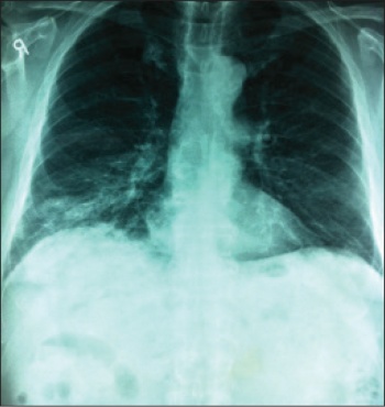 Figure 1. Initial CXR on presentation(click to enlarge)


