Rare entity of paraurethral cyst in newborn infant
A case on the diagnosis and management of an interlabial mass in a newborn female infant
Dr Muhammad Jawad, Locum Consultant Paediatrician, Wexford General Hospital, Wexford and Dr Frances Neenan, Consultant Paediatrician, Portiuncula Hospital, Ballinasloe
A female infant was born at term following a spontaneous vaginal delivery to a 40-year-old Irish woman. The pregnancy was uneventful and antenatal foetal anomaly scan did not reveal any structural abnormality. The infant was vigorous at birth and APGAR scores were good (91 and 95 minutes) with a birth weight of 3.4kg.
She had a normal systemic physical examination except for an oval shaped mass in her vulval area measuring 2 x 1cm (see Figure 1).
Figure 1. Oval shaped mass in vulval area measuring 2 x 1cm(click to enlarge)
The mass was yellow/white in colour, thin walled, shiny and had a superficial vascularity in the anterior area. It was firm in consistency, obscuring the vaginal opening and pushing the urethral opening to the left lateral side of the visible swelling.
She had a good urinary stream noted by nursing staff. An ultrasound examination of the mass revealed a homogenous, diffusely echogenic structure in the interlabial area with no connection to the urinary system and no Doppler evidence of intralesional vascularity suggestive of a cystic lesion.
A diagnosis of paraurethral cyst was made based on the unique features of the mass. Her case was discussed with the paediatric surgical team and a diagnosis of paraurethral cyst was agreed. She was reviewed by the paediatric surgeon in outpatients on day 10 of life and the mass was noted to be regressing in size.
A conservative treatment approach of watchful waiting was adopted and further follow up was planned in surgical outpatients.
A follow up visit at paediatric outpatient clinic at two and half months revealed complete resolution of this cystic mass.
Discussion
Paraurethral cysts arise from paraurethral glands, and open into the female urethra by ducts. There are six to 30 ducts, the largest two called the Skene’s ducts. The aetiology of development of paraurethral cysts is unknown. Two postulated theories mentioned in the literature are:
The possibility of dislocated uroepithelium from the urogenital sinus leading to obstruction of glandular ducts and the developmental of paraurethral cysts1
Maternally acquired hormones stimulate glandular secretions leading to cyst formation.1
Incidence
Paraurethral cysts are a rare entity in newborn females. In old literature, reported incidence was one in 7,246 but recent studies have reported one in 1,038.2
Only 54 cases have been reported so far in the English literature, mainly in surgical and radiological journals.2,4
Differential diagnosis
The differential diagnosis of an interlabial mass includes:
Hydrometrocolpos
Gartner’s duct cyst
Prolapsed urethra
Prolapsed ectopic ureterocele
Rhabdomyosarcoma of the vagina.
The appearance of the mass, its position relative to the urethral opening, the pattern of the urinary stream, and the child’s age and race help to determine the diagnosis.
A hydrometrocolpos appears as a bulging mass at the vaginal opening, pearly grey in colour and has a normally positioned urethral meatus.3
A urethral prolapse tends to occur more in girls of African origin and rarely in infants. It appears as red/dark blue in colour (depending on vascular compromise), oedematous, often haemorrhagic mass and encircles the urethral opening leading to the central urinary stream.3
A prolapsed ureterocele almost always occurs in Caucasian girls, light pink to dark purple in colour, without an identified urethral meatus and allows circumferential urinary flow unless it causes complete obstruction.3
Rhabdomyosarcoma of the vagina presents as a mass protruding through the vaginal orifice, pearly grey in colour and resembles a cluster of grapes.3
Ultrasonography and urological examination can differentiate between the above mentioned interlabial masses. Perineal ultrasonography has been shown to diagnose paraurethral cysts without detailed invasive examination of the urogenital system.5
Management
There has been a lot of disagreement regarding treatment of paraurethral cysts in literature. Excision and marsupilisation of the mass was mentioned in earlier studies.4,6 However, more recent studies have revealed that the paraurethral cysts will regress spontaneously.
Fujimoto et al followed five patients with paraurethral cysts conservatively for a five year study period, all cases regressed spontaneously in 76 to 304 days.2
Conclusion
Paraurethral cysts are a relatively uncommon finding. A thorough examination of the urogenital area should be an essential part of the newborn physical examination to find such masses. Physical examination alone will be sufficient to diagnose paraurethral cysts, however further investigations can be performed if uncertainties exist.
Management remains controversial and further studies are required to find the best treatment approach.
References
Soyer T, Aydemir E, Atmaca E. Paraurethral cysts in female newborns: role of maternal oestrogens. J Paediatr Adolesc Gynecol 2007; 20: 249-251
Fujimoto T, Suwa T, Ishii N et al. Paraurethral cyst in female newborn: is surgery always advocated? J Pediatr Surg 2007; 42:400
Nussbaum AR, Lebowitz RL. Interlabial masses in little girls: review and imaging recommendations. Am J Roentgenol 1983; 141:65
Blaivas JG, Pais VM, Retik AB. Paraurethral cysts in female neonate. Urology 1976; 7: 504-507
Breysem L, Rayyan M, Bogaret G, Vanhole C, Smet MH. High resolution parineal ultrasound of a paraurethral cyst in a neonate. Eur Radiol 2008; 18:2,701-2: 703
Ceylan H, Ozokutan BH, Karak ok M et al. Paraurethral cyst: is conservative management always appropriate? Eur J Pediatr Surg 2002; 12: 212-214
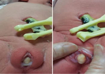 Figure 1. Oval shaped mass in vulval area measuring 2 x 1cm(click to enlarge)