New developments in imaging technology are now integral in the diagnosis and follow up of many serious ophthalmic conditions
Dr Rory Murphy, Ophthalmology Trainee, Cork University Hospital, Cork, Dr Treasa Murphy, Basic Specialist Trainee in Ophthalmology, Royal Victoria Eye and Ear Hospital, Dublin, Dublin and Dr Yvonne Delaney, Dean of Postgraduate Education, Irish College of Ophthalmologists, Dublin
By allowing light of different wave lengths to enter via the pupil, the eye is the ideal organ to take advantage of new developments in imaging technology. The most important of these advances allows unprecedented detailed imaging of the anterior and posterior segments of the eye, especially the retina, the retinal vasculature and the optic nerve.
Optical coherence tomography (OCT)
Optical coherence tomography (OCT) is a non-contact, non-invasive imaging technique. Invented in 1991, it exploits the property of low coherence laser interferometry and optical back scattered light to derive depth information of various retinal structures.1 This is achieved by comparing the time difference in reflected light from the retina at various depths with a reference ‘standard’. Differences between the reflected light and the reference standard provide structural information in the form of a reflectivity profile called an A-scan. By laterally combining a series of these axial depth scans (A-scan), a cross-sectional tomograph (B-scan) of the retina or optic nerve can be generated.2 The resolving power of this new technology, at about 10 micrometres vertically and 20 micrometres horizontally, is unprecedented with its nearest rival of ultrasound achieving a resolution of 100 microns or less.3
OCT has become integral to the diagnosis and treatment of retinal and other ocular pathologies. The past decade has seen an even greater enhancement of OCT techniques in terms of micrometre resolution and cross-sectional as well as three-dimensional imaging capability. This has led to the visualisation of previously unknown ocular pathology, improved diagnostic techniques, and the utilisation of OCT as an evaluation tool for new pharmacological and surgical treatments.
(click to enlarge)
Swept-source OCT (SS-OCT)
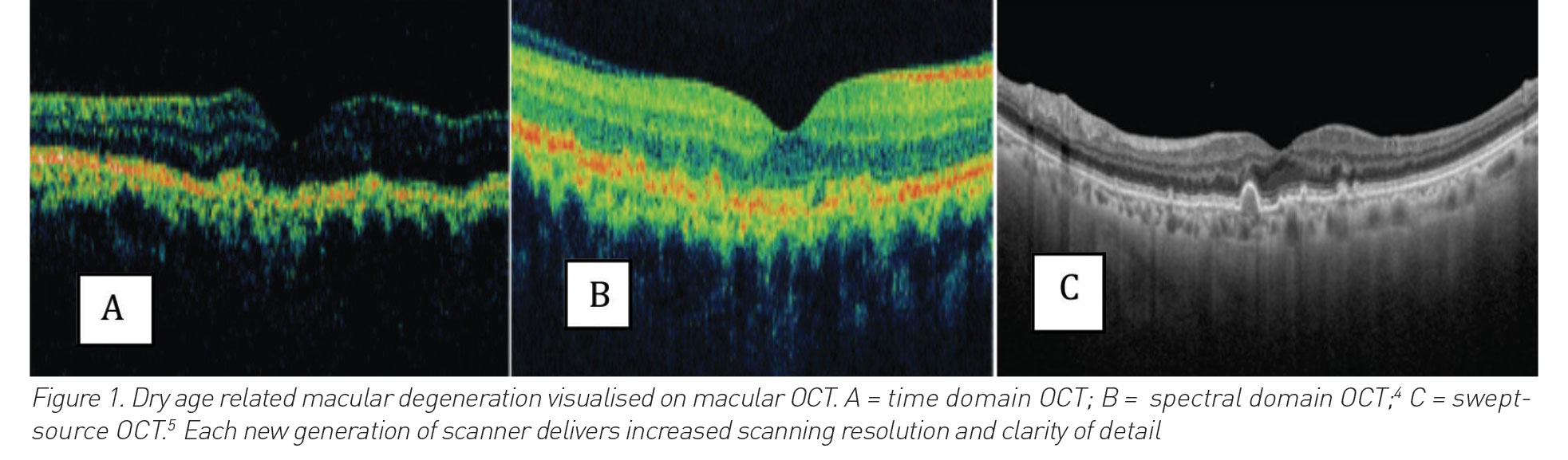
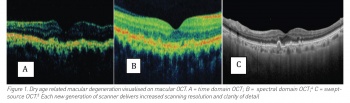
Swept-source OCT (SS-OCT) is the most advanced form of OCT. It represents the third generation of OCT development, following on from time-domain (TD) and spectral domain (SD) OCT (see Figure 1). SS-OCT differs from its predecessors by utilising a tuneable swept laser as the light source and a single photodiode as the detector. This allows it to operate faster, scanning wider and deeper images of the retina. The speed at which the SS-OCT functions has been groundbreaking, generating 100,000-400,000 A-scan images per second, which is double the speed of its spectral domain predecessor.3
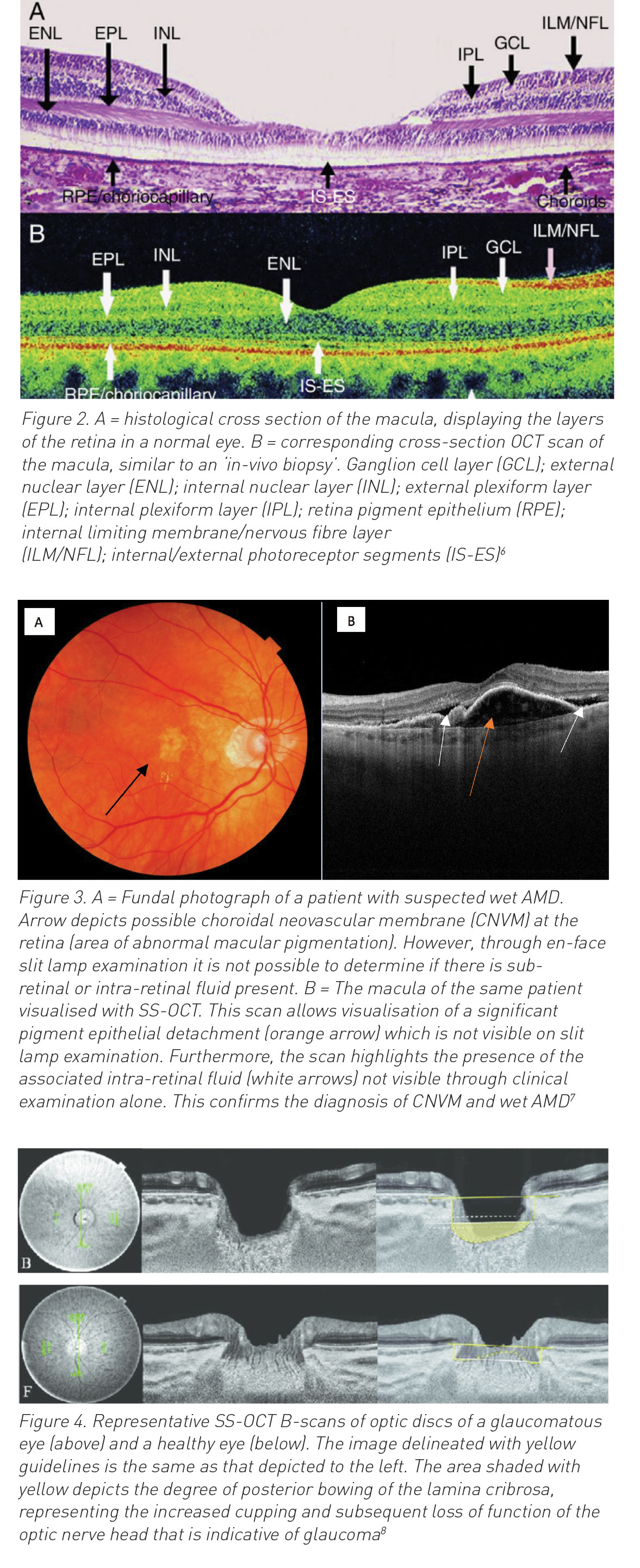
The resolution of the SS-OCT is also unprecedented. It represents the closest technological advancement to an in-vivo biopsy of the retina and allows detailed visualisation of microscopic retinal structures which may be as thin as 2-3µm (see Figure 2).
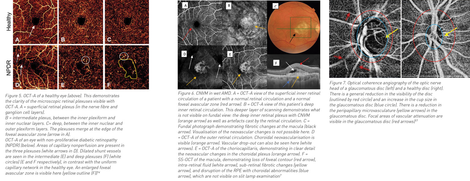
Furthermore, SS-OCT enables deeper visualisation of pathology under the retina and under the retina pigment epithelium (RPE), pathology that was previously not visible on the earlier versions of TD and SD OCT. This ability is especially relevant in macular diseases such as age-related macular degeneration (AMD) where pathological blood vessels may lie under the RPE (see Figure 3).
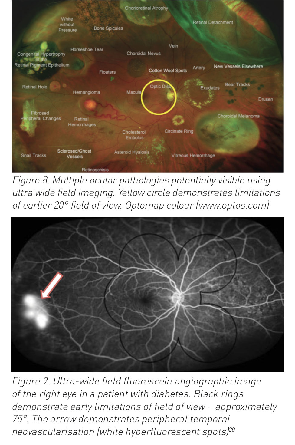
SS-OCT technology has additionally contributed to the understanding of other common eye diseases such as glaucoma where it can be utilised to monitor glaucoma progression through measurement of the depth of the retinal nerve fibre layer and ganglion cell layer of the retina, in slices that are less than 3µm thick (see Figure 4).
(click to enlarge)
Optical coherence angiography (OCTA)
OCTA algorithms detect vascular flow information by generating images based on the motion signal between repeated OCT B-scans at the same position. This motion signal, which reflects subtle differences between the B-scans, is mostly due to the movement of erythrocytes within blood vessels.9,10 This enables OCT angiography to create detailed non-invasive images of the retinal and choroidal vasculature with visualisation of vessels as small as 2-3µm in diameter. Prior to OCTA, fundal fluorescein angiography (FFA) was the only technique available to evaluate the retinal circulation. While still extremely useful in the evaluation of retinal vascular disease, its drawbacks are considerable. It is labour intensive, requires the intravenous injection of a fluorescein dye (with rare but significant side effects) and it does not allow the direct visualisation of the underlying sub-retinal or choroidal vasculature.11
OCTA allows visualisation of pathology such as microaneurysms and neovascularisation, signs that can be detected clinically on fundoscopy but in detail that was previously unobtainable.12 More importantly OCTA allows imaging of aspects of the retinal and choroidal circulation which cannot be visualised on clinical examination. Three different levels of the circulation can be imaged – the retina, under the retina and under the RPE (see Figure 5).13
Furthermore, OCTA enables a different approach to visualising pathologies that were previously unseen on clinical examination and fluorescein angiography, such as occult choroidal neovascular membranes (see Figure 6)15 and vascular attenuation of the glaucomatous optic nerve (see Figure 7).16
(click to enlarge)
Ultra-wide field imaging
Imaging the fundus was made possible in 1926 by Zeiss and Nordensen when they created a fundal camera with a 20° field of capture. Essentially this captured a small area and was limited to viewing the disc, macula and posterior pole (yellow circle on Figure 8). Since then, we have witnessed an evolution into highly sophisticated non-contact scanning laser camera systems, such as the Optos camera capturing 200° ultra-wide field (UWF), high-resolution images, covering approximately 82% of the retinal area while retaining remarkable clarity.18 This has translated into outstanding diagnostic and prognostic capabilities for the treating ophthalmologist. Some potentially visible pathology seen using the Optos camera are displayed in Figure 8, in an overlay image of multiple possible ocular comorbidities.
(click to enlarge)
UWF imaging and fundal fluorescence angiography (FFA)
The advantages of UWF imaging are not limited to colour photography, and are now routinely combined with the well-established fundus fluorescein angiography (FFA). Invented by two medical students, Novotny and Alvis in 1961, FFA has proven to be of huge benefit in diagnosing vascular defects across a myriad of pathologies.19 Peripheral non-perfusion in people with diabetes is elegantly depicted by the combined use of UWF imaging and FFA (see Figure 9), helping identify those individuals at risk for blinding proliferative disease.
Figure 9 shows a right-sided FFA of a patient with diabetes, captured with wide field imaging. Until the advent of UWF, the area captured would have been limited to within the black rings, which is essentially seven standard photographs overlaid to show 75°. The neovascularisation (arrow) is outside this region, and so would previously have been missed, yet is clearly visible in the UWF image (200°) and flags the need for urgent sight -saving laser treatment. A recent study comparing UWF imaging to the previous standard imaging, showed a 3.9‑fold increase in diagnosing non-perfusion (p < 0.001), and a 1.9-fold increase in diagnosing neovascularisation (p < 0.0036).20 With an increasing prevalence of diabetes, and huge potential burden of diabetic eye disease, UWF imaging with FFA has been instrumental in capturing the full cohort of at-risk individuals, improving screening, translating to reduction in the prevalence of blindness. A three-fold increased risk in diabetic retinopathy progression can be established in the presence of predominantly peripheral lesions, and a nearly five-fold increased risk of proliferative retinopathy, signalling those requiring sight-saving treatment in the form of pan retinal photocoagulation.21
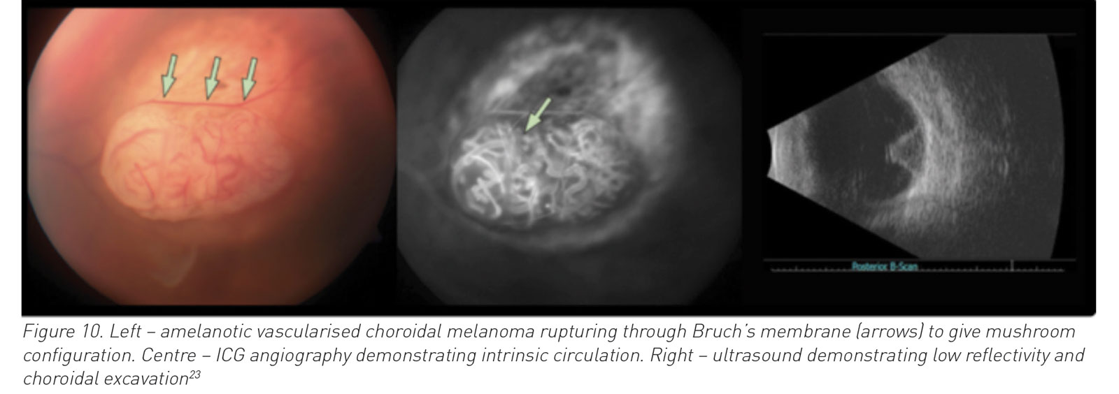
Furthermore, any vascular event such as retinal vein occlusions (RVO), arterial occlusions (RAO), retinal vasculitis and posterior uveitis, infer risk of ischaemia and subsequent neovascularisation leading to blindness.22 The management of many of these individuals now relies heavily on UWF imaging with FFA. Management in the form of appropriate sectoral, or pan retinal photocoagulation can help avoid secondary complications and preserve vision. Choroidal naevi or ocular tumours in the periphery can also be detected on UWF colour photography, (see Figure 8) and monitored for suspicious signs or progression indicative of malignant disease. Further imaging modalities in suspected tumours include ICG angiography to look for double circulation signs, and B-scan ultra sound scanning to assess degree of echoic reflectivity (see Figure 10).
(click to enlarge)
(click to enlarge)
Fundal autofluorescence (FAF)
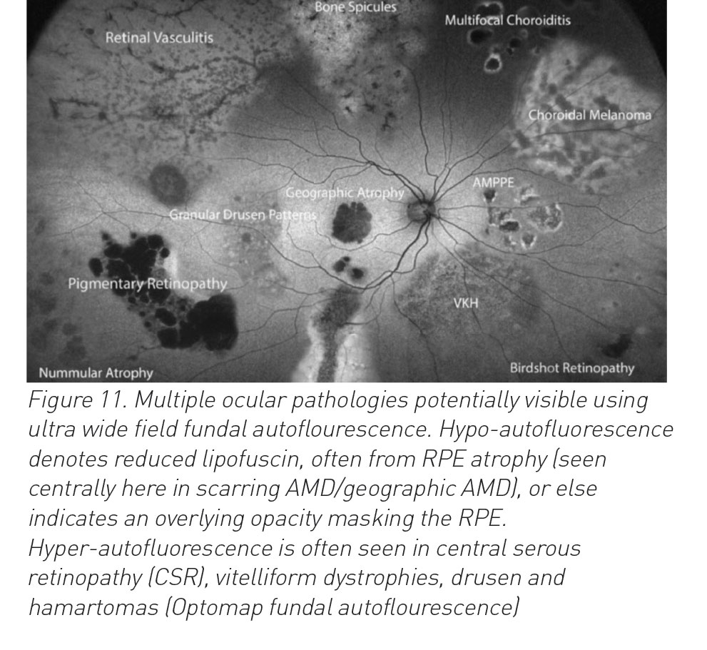
Fundal autofluorescence (FAF) is another non-invasive imaging modality, which essentially forms a lipofuscin density map and assesses the health of the retinal pigment epithelial layer (RPE), of particular use in dry forms of age-related macular degeneration. Lipofuscin, being the predominant fluorophore of the RPE, absorbs and emits light at a specific wavelength and has been in use for retinal photography since the 1980s.24 Its use is expanding but includes pathologies such as macular dystrophies, retinitis pigmentosa, white dot syndrome and drug toxicities. UWF FAF has also been shown to help in the management of rhegmatogenous retinal detachments25 and also to detect distinct peripheral abnormalities often seen in age-related macular degeneration.26
Anterior segment OCT
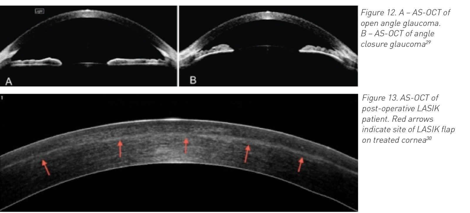
The development of anterior segment OCT (AS-OCT) has enabled significant imaging of the anatomy of the anterior ocular segment. It operates at 1310nm wavelength to acquire superior images of the conjunctiva, cornea, iris, anterior chamber and its angles, including the trabecular meshwork and Schlemm’s canal.27 These were previously only visible clinically through gonioscopy lenses, held in contact with the patient’s globe. Before the development of AS-OCT, there was no technology enabling imaging of the angle of the eye. This imaging is now integral in the diagnosis and follow up of serious conditions, such as acute angle closure glaucoma28(see Figure 12).
Additionally, AS-OCT is detailed enough to enable visualisation of the microscopic corneal stroma and flap in treatment planning for corneal refractive procedures such as LASIK (laser in si tu keratotomy )30(see Figure 13). Its uses are constantly progressing with current investigation into the technology’s suitability in assessing tear film in dry eyes, contact lens fit, and measuring corneal power in post-operative LASIK patients awaiting cataract surgery.
(click to enlarge)
Future prospects
Future prospects of retinal imaging are promising, particularly in the area of telemedicine which may yet play a large role in reducing global preventable blindness. Non-physician operators in resource-poor areas can be trained to acquire images and their findings evaluated remotely by physicians, for example in diabetic retinopathy and retinopathy of prematurity where specialist clinicians might not be on site.31 Furthermore, developing countries with limited infrastructure and access to ophthalmic review may benefit from mobile phone and hand-held retinal photography.20,32
References
Gabriele ML, Wollstein G, Ishikawa H et al. Optical Coherence Tomography: History, Current Status, and Laboratory Work. Investigative Ophthalmology & Visual Science 2011; 52(5):2425-36
Lee MR et al. Optical coherence tomography for ophthalmic imaging: new technique delivers micron-scale resolution. IEEE Engineering in Medicine and Biology Magazine, 1995; 14(1):66-67. doi: 10.1109/51.34075
Potsaid B, Baumann B, Huang D et al. Ultrahigh speed 1050nm swept source/Fourier domain OCT retinal and anterior segment imaging at 100,000 to 400,000 axial scans per second. Optics express 2010; 18(19):20029-20048
Gerson R. Spectral domain: the land after time. Review of Optometry 2008; 145(08)
Ghadkar A, Kumar V et al. Swept source ocular coherence tomography: visualising vitreous, retina and choroid simultaneously. Delhi J Ophthalmol 2017; doi: 10.7869/djo.269
Lasave AF. Archives of the Spanish Society of Ophthalmology. Current interpretation of optical coherence tomography in the posterior pole. 2016; 91:3-9. doi: 10.1016 / j.oftale.2015.12.013
Ruiz Moreno JM. Topcon Triton DRI Swept source OCT. Universidad Castilla la Mancha, Albacete, Spain
Akil H et al, Optical Coherence Tomography Angiography of the Optic Disc; an Overview. J Ophthalmic Vision Research 2017(Jan-Mar); 12(1): 98-105. doi: 10.4103/2008-322X.200162
Kim DY, Fingler J, Zawadzki RJ et al. Optical Imaging of the chorioretinal vasculature in the living human eye. Proc Natl Acad Sci 2013; 110:14354-9
Spaide RF, Klancnik JM, Cooney MJ. Retinal vascular layers imaged by fluorescein angiography and optical coherence tomography angiography. JAMA Ophthalmol 2014; E1-6. doi:10.1001/jamaophthalmol.2014.3616
Novotny HR, Alvis DL. A method of photographing fluorescence in circulating blood in the human retina. Circulation 1961; 24:82-6
Choi W, Waheed NK, Moult EM et al. Ultrahigh Speed swept source optical coherence tomography angiography of retinal and choriocapillaris alterations in diabetic patients with and without retinopathy. Retina 2017; 37(1):11-21
De Carlo T, Romano F et al. A review of optical coherence tomography angiography (OCTA). Int J Retina Vitreous 2015; 1(5) doi:10.1186/s40942-015-0005-8
Gao SS, Jia Y, Zhang M et al. Optical Coherence Tomography Angiography. Invest Ophthalmol Vis Sci 2016; 57(9): OCT27-OCT36. doi: 10.1167/iovs.15-19043
Bailey S, Jia Y, Huang D et al. Detection of occult choroidal neovascularization in age-related macular degeneration with optical coherence tomography angiography. Invest Ophthalmol Vis Sci 2015; 56(7 ):3329
Jia Y, Wei E, Wang X et al. Optical coherence tomography angiography of optic disc perfusion in glaucoma. Ophthalmology 2014; 121(7):1322-1332. doi: 10.1016/j.ophtha.2014.01.021
Hanadan A, Alfredo S et al. Optical coherence tomography angiography of the optic disc; an overview. J Ophthalmic Vision Research 2017; 12(1):98-105
Witmer MT, Parlitsis G, Patel S, Kiss S. Comparison of ultra-widefield fluorescein angiography with the Heidelberg Spectralisnoncontact ultra-widefield module versus the Optos Optomap. Clin Ophthalmol 2013; 7:389-94
Novotny HR, Alvis DL. A method of photographing fluorescence in circulating blood in the human retina. Circulation 1961;24:82-6
Wessel MM, Aaker GD, Parlitsis G et al. Ultra-wide-field angiography improves the detection and classification of diabetic retinopathy. Retina 2012; 32(4):785-91
Sim DA, Keane PA, Rajendram R et al. Patterns of peripheral retinal and central macula ischemia in diabetic retinopathy as evaluated by ultra-widefield fluorescein angiography. Am J Ophthalmol 2014;158(1):144-53.e1
Leder HA, Campbell JP, Sepah YJ et al. Ultra-wide-field retinal imaging in the management of non-infectious retinal vasculitis. J Ophthalmic Inflamm Infect 2013; 3(1):30
Delori FC, Dorey CK, Staurenghi G et al. In vivo fluorescence of the ocular fundus exhibits retinal pigment epithelium lipofuscin characteristics. Invest Ophthalmol Vis Sci 1995; 36(3):718-29
Witmer MT, Cho M, Favarone G et al. Ultra-wide-field autofluorescence imaging in non-traumatic rhegmatogenous retinal detachment. Eye (Lond) 2012; 26(9):1209-16
Tan CS, Heussen F, Sadda SR. Peripheral autofluorescence and clinical findings in neovascular and non-neovascular age-related macular degeneration. Ophthalmology 2013; 120(6):1271-7
Ramos JLB, Li Y, Huang D. Clinical and research applications of anterior segment optical coherence tomography – a review. Clinical & Experimental Ophthalmology 2009; 37(1):81-9
Radhakrishnan S, Goldsmith J, Huang D t al. Comparison of optical coherence tomography and ultrasound biomicroscopy for detection of narrow anterior chamber angles. Archives of Ophthalmology (Chicago, Ill : 1960). 2005; 123(8):1053-9
Kent C. Making the Most of Anterior Segment OCT. Review of Ophthalmology 2011. Image courstesy of Prof Joel S. Schuman, University of Pittsburg
Li Y, Netto MV, Shekhar R, Krueger RR, Huang D. A longitudinal study of LASIK flap and stromal thickness with high-speed optical coherence tomography. Ophthalmology 2007; 114(6):1124-32
Fijalkowski N, Zheng LL, Henderson MT et al. Stanford University Network for Diagnosis of Retinopathy of Prematurity (SUNDROP): five years of screening with telemedicine. Ophthalmic Surg Lasers Imaging Retina 2014; 45(2):106-13
Panwar N, Huang P, Lee J et al. Fundus photography in the 21st century - a review of recent technological advances and their implications for worldwide healthcare. Telemed J E Health 2016; 22(3):198-208
 (click to enlarge)