A 23-year-old woman had two previous pregnancies which were delivered by Caesarean sections, and were pre-term deliveries (34 weeks, 35 weeks) and the third was delivered at term. She gave a history of abuse when she was a child by a close relative and all her children are on the child protection register. At 27 weeks gestation she was admitted with mild back pain, investigations were normal and the midstream specimen of urine (MSSU) was negative, steroid injections for foetal lung maturity were given. The pain had settled the next day and she was allowed to go home. At 31 weeks gestation she was re-admitted with back pain and tightness, her vital signs were normal, no contractions were felt, cardiotocography (CTG) was reassuring with no uterine activity, and the patient was kept for observation.
Five hours later she started to complain of scar tenderness and was distressed with the pain. On examination vital signs were normal, abdomen soft with localised tenderness to the site of the previous Caesarean scar, vaginal examination showed 1.5cm long closed cervix posterior no leakage or bleeding, CTG showed a normal trace with no uterine activity. Intravenous fluid started, 50mg pethidine did not improve the pain, the blood pressure went from 117/80 to 86/34 and remained low for two hours and 40 minutes. The patient was insisting that she was in severe pain, the impression was that it could be a wound dehiscence.
A decision was made to perform an emergency Caesarean section and the patient was transferred to the operating theatre. Only then did her symptoms fade and she looked more comfortable and stopped complaining of any pain. Moreover she was exceptionally pleasant to the staff.
The Caesarean section was carried out under a spinal anaesthesia and the neonatal staff were present. The lower uterine segment was well formed with no rupture or scar dehiscence identified. There were mild adhesions.
The outcome was a male baby of 1,750g with Apgar’s of 1/10 and 5/10, blood loss was average, abdomen wall closed in layers, but post-operatively all vital signs were normal. The baby was transferred to the tertiary neonatal unit on the second postnatal day with his mother.
Figure 1(click to enlarge)
Figure 2(click to enlarge)
Discussion
Experience associated with childhood sexual abuse may affect the incidence and the outcome of adolescent childbearing, identification and treatment of previously abused adolescent prenatal patients may break this vicious intergenerational cycle of violence.1
This patient who had undergone three previous Caesarean sections,was known to health and social services, and had been complaining of localised tenderness at the Caesarean sections scar. She was not responding to pethidine, but CTG was reassuring. Blood pressure dropped; however, when the decision for a Caesarean section was made and the patient as previously stated was transferred to the operating theatre, she looked comfortable and stopped complaining of any pain. The disappearance of pain at this stage make the attending medical staff uncomfortable as this may indicate uterine scar gaping or rupture.
Uterine rupture
Uterine rupture in pregnancy is a rare complication, even in high-risk subgroups; the overall incidence of uterine rupture is low. Twenty peer-reviewed publications between 1976-2009 reported 1,864 uterine ruptures among 2,863,330 pregnant women, yielding an overall rate of one in 1,536 pregnancies (0.07%).2 A woman who has had more than one Caesarean section with a low horizontal incision may have a slightly higher risk of rupture.3
Uterine rupture during pregnancy is a catastrophic complication that frequently results in life-threatening maternal and foetal compromise. Full thickness separation of the uterine wall and the overlying serosa will lead to significant uterine bleeding and foetal distress. From the time of diagnosis to delivery only 10-37 minutes are available before clinically significant foetal morbidity becomes inevitable.2
The foetal morbidity occurs as a result of catastrophic haemorrhage, foetal anoxia or both. Protrusions of the foetus or placenta or both into the abdominal cavity necessitate the need for prompt Caesarean delivery and sometimes the damage to the uterus is beyond repair and a hysterectomy is required. One study reported better outcomes when the response time was 18 minutes or less.3
However, most guidelines allow for a maximum response time of 30 minutes. The longer it takes to respond to a uterine rupture the more likely it is that the baby and/or the placenta can be pushed through the uterine wall and into the mother’s abdominal cavity.3
Uterine scar dehiscence, on the other hand, is a more common event which constitutes separation of a pre-existing scar and that does not disrupt the overlying visceral peritoneum (uterine serosa) and that does not significantly bleed from its edges and it seldom results in major maternal or foetal complications.2
Premonitory signs of uterine rupture include foetal heart rate abnormalities, loss of uterine contractility, abdominal pain, recession of the presenting foetal part andvaginal bleeding; all these signs are inconsistent.4
There is no reliable method to predict which uterus will have a rupture and these initial signs and symptoms of uterine rupture are typically non-specific, which makes diagnosis difficult and sometimes delays definitive therapy. Moreover, uterine rupture cannot be ‘predicted’ with either individual or combinations of clinical factors, this has important clinical and medico-legal implications.5
A study has been done for ‘prediction’ of complete uterine rupture by sonographic evaluation of the lower uterine segment and the conclusion of the study showed that full lower uterine segment thickness of < 2.3mm is associated with a higher risk of complete uterine rupture.6 However, because of the short time available to diagnose uterine rupture before the onset of irreversible physiological damage to the foetus, time-consuming diagnostic methods and sophisticated imaging modalities have only limited place. Therefore, uterine rupture is most appropriately ‘diagnosed’ on the basis of standard signs and symptoms.4
An abnormal pattern in foetal heart rate was the first manifestation of uterine rupture and was found in 87% of patients and in fact is the most common finding associated with uterine rupture 79% when cases involve the extrusion of the placenta and foetus into the abdominal cavity, prolonged decelerations in foetal heart rate invariably occurred. Sudden or atypical abdominal pain is less common than foetal heart rate decelerations or bradycardia, in nine studies from 1980-2002, abdominal pain occurred in 13-60% of cases of uterine rupture.
In a review of 10,967 patients undergoing a trial of labour, only 22% of complete uterine ruptures presented with abdominal pain.
Moreover, in a study by Bujold and Gauthier, abdominal pain was the first sign of rupture in only 5% of patients and occurred in women who developed uterine rupture without epidural analgesia but not in women who received an epidural block thus abdominal pain may be an unreliable and uncommon sign of uterine rupture. The loss of the uterine contraction may not be reliable either as with uterine rupture, labour sometimes continues, without loss of uterine tone or amplitude of contractions.4
Delivery at 31 weeks gestation is not without its complications. Prematurity and its complications are the underlying reasons for the higher rate of infant mortality and morbidity.7 Even later in life when IQ tested, eg. at 20 years of age, approximately 42% of very low birth-weight infants have been found to have borderline IQ scores (70-84) and 7% had subnormal IQ scores (less than 70), compared to 31% and 2% respectively in normal birth-weight infants.8
Recent follow-up studies have also revealed that these infants may demonstrate more subtle impairments such as learning disabilities, impaired social skills, and behavioural problems, particularly attention deficit-hyperactivity disorder.9 Data describing significant long-term morbidities and neurodevelopmental outcomes based upon birth weight and gestational age at delivery, the early identification of individuals at risk for these impairments remains an ongoing challenge for physicians.
Acknowledging our limited capability to predict which infants will be most severely affected is crucial for effective and honest communication with families.10
Conclusion
Uterine rupture in pregnancy is a rare and catastrophic complication that frequently results in maternal life-threatening and foetal compromise. Time-consuming diagnostic methods and sophisticated imaging modalities have limited place. The inconsistency of premonitory signs and the short time for instituting definitive therapeutic action make uterine rupture a fearful event. Caesarean section was performed, at 31 weeks gestation in this case and this was based on the history of pre-term delivery, persistence of pain at the scar area after analgesia and changes in blood pressure, causing long-term infant morbidities based upon birth weight and gestational age at delivery. The early identification of individuals at risk remains an ongoing challenge for physicians.
History of childhood sexual abuse lowers the pain threshold and may affect the incidence and the outcome of adolescent childbearing. Therefore, identification and treatment of previous abuse may break this vicious intergenerational cycle of violence and such history is needed to be highlight at the patient’s notes which may affect the management by the attending medical staff.
In this case such history was not highlighted, which may have weighed in the final management plan.
References
Stevens-Simon C, Reichert S. Sexual abuse, adolescent pregnancy, and child abuse. Arch Pediatr Adolesc Med 1994; 148 (1): 23-27
Nahum GG, Chelmow D. http://emedicine.medscape.com/article/275854-overview May 12, 2010
Jukelevics N. http://www.obgyn.net/pregnancy-birth/article/7273
Nahum GG, Chelmow D. Uterine rupture in pregnancy, signs and symptoms of uterine rupture during pregnancy, 2010. http://emedicine.medscape.com/article/275854-overview#aw2aab6b6
Macones GA, Cahill AG, Stamilio DM et al. Can uterine rupture in patients attempting vaginal birth after cesarean delivery be predicted? Am J Obstet Gynecol 2006; 195(4): 1148-1152
Bujold E, Jastrow N, Simoneau J et al. Prediction of complete uterine rupture by sonographic evaluation of the lower uterine segment. Am J Obstet Gynecol 2009; 201(3): 320. e1-6
Mandy GT. Short-term complications of the premature infant, updated: September 30, 2010, http://www.uptodate.com/contents/short-term-complications-of-the-premature-infant?source=search_result&selectedTitle=20%7E150
Hack M, Flannery DJ, Schluchter M et al. Outcomes in young adulthood for very-low-birth-weight infants. N Engl J Med 2002; 346(3): 149-157
Hack M. Young adult outcomes of very-low-birth-weight children. Semin Fetal Neonatal Med 2006; 11(2): 127-137
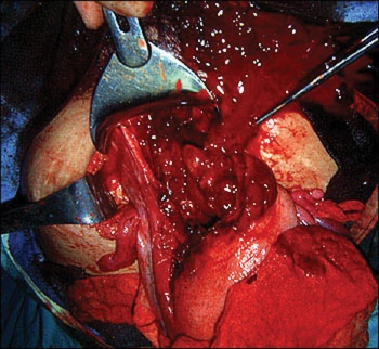 Figure 1(click to enlarge)
