SURGERY

UROLOGY

Understanding bladder voiding dysfunction

To understand the concept of bladder dysfunction, it is important to remember the mechanism by which the bladder develops, and its relationship to the nervous system and adjacent structures

Mr Fardod O'Kelly, Specialist Registrar in Urology, Department of Urological & Reconstructive Surgery, Mater Misericordiae University Hospital, Dublin and Mr Kiaran O'Malley, Consultant Urologist, Department of Urological & Reconstructive Surgery, Mater Misericordiae University Hospital, Dublin

March 1, 2015

Article
Similar articles
  • The bladder is an incredibly complex organ, which develops from the anterior cloaca, a structure that has exquisite evolutionary conservatism, as it performs a vital task across phyla of species from birds to reptiles to mammals, with only small adjustments across species. Interestingly, humans are one of the only species to actually suffer from bladder dysfunction, which demonstrates the obvious and complex links with high order centres.

    Bladder embryology

    During foetal development, the terminal part of the hindgut ends in the cloaca, which is an endoderm-lined chamber that contacts the surface ectoderm at the cloacal membrane and communicates with the allantois, which is a membranous sac that extends into the umbilicus alongside the vitelline duct. The cloaca is then divided by the urorectal septum with the dorsal (inferior) portion developing into the rectum and anal canal, and the ventral (superior) portion developing into the bladder and urogenital sinus, which will give rise to the bladder and lower urogenital tracts (prostatic and penile urethrae in males; urethra and lower vagina in females). As the bladder grows and expands, the distal ends of the mesonephric ducts are absorbed into the wall of the bladder as the trigone. Malformations during development (aside from a condition involving failed reinforcement of the cloacal membrane by mesoderm, such as bladder exstrophy) can lead to a number of conditions such as abnormal ureter attachments, urachal fistulas, sinuses and cysts.

    Bladder anatomy and physiology

    The bladder acts not only as a temporary store for urine before its coordinated expulsion through the urethra at an appropriate time and place, but also as an autocrine organ involved in its own regulation. 

    The bladder is the most anterior element of the pelvic viscera. It is situated in the pelvic cavity when empty, but expands superiorly into the abdominal cavity when full. The urinary bladder is abdominal at birth, positioned at the extraperitoneal area of the lower abdominal wall. At around the fifth or sixth year of age, the bladder gradually descends into the area of the true (minor) pelvis. It contains four main parts: the apex, the base, and the superior and inferolateral surfaces. The mucosal lining on the base of the bladder is smooth and firmly attached to the underlying smooth muscle coat of the wall – unlike elsewhere in the bladder where the mucosa is folded and loosely attached to the wall. The smooth triangular area between the openings of the ureters and urethra on the inside of the bladder is known as the trigone. 

    The vesicoureteric junctions is found as the ureter approaches the bladder. At 2cm to 3cm from the bladder, a fibromuscular sheath (of Waldeyer) extends longitudinally over the ureter and follows it to the trigone. The ureter pierces the bladder wall obliquely, travels 1.5cm to 2cm, and terminates at the ureteral orifice. As it passes through a hiatus in the detrusor (intramural ureter), it is compressed and narrows considerably. The intravesical portion of the ureter lies beneath the urothelium, it is backed by a strong plate of detrusor muscle. With bladder filling, this arrangement is thought to result in passive occlusion of the ureter, like a flap valve. 

    This anatomic arrangement helps prevent reflux during bladder filling by fixing and applying tension to the ureteral orifice. As the bladder fills, its lateral wall telescopes outward on the ureter, thereby increasing intravesical ureteral length. Vesicoureteral reflux is thought to result from insufficient submucosal ureteral length and poor detrusor backing. Chronic increases in intravesical pressure resulting from bladder outlet obstruction can cause herniation of the bladder mucosa through the weakest point of the hiatus above the ureter and produce a ‘Hutch diverticulum’ and reflux. 

    One of the reasons that bladders can work so efficiently irrespective of the stimulus, is as a result of transitional cell epithelium, which is waterproof and distensible, and allows bladder filling without a concomitant rise in pressure (compliance). Bladder filling from the excretion of urine by the kidneys occurs via the ureters. The walls of the ureters contain smooth muscle arranged in spiral, longitudinal and circular bundles, but distinct layers of muscle are not seen. Regular peristaltic contractions occurring one to five times per minute move the urine from the renal pelvis to the bladder, where it enters in spurts synchronous with each peristaltic wave. The ureters pass obliquely through the bladder wall and, although there are no ureteral sphincters as such, the oblique passage tends to keep the ureters closed except during peristaltic waves, preventing reflux of urine from the bladder. An adult bladder should hold somewhere between 400ml and 600ml, whereas children require a separate calculation: Expected bladder capacity = [30 + (age in years x 30)] ml.

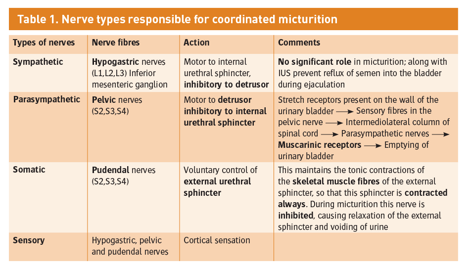
    During bladder emptying, contraction of the circular muscle, which is called the detrusor muscle, is mainly responsible. Muscle bundles pass on either side of the urethra and these fibres are sometimes called the internal urethral sphincter (smooth muscle), although they do not encircle the urethra. Farther along the urethra is a sphincter of skeletal muscle, the sphincter of the membranous urethra, external urethral sphincter. This needs to happen in an extremely coordinated manner as described below, and micturition is therefore fundamentally a spinal reflex, facilitated and inhibited by higher brain centres and, like defecation, subject to voluntary facilitation and inhibition (see Table 1).

    Bladder filling and a desire to void stimulates afferent impulses to the sacral micturition centre in the spinal cord which allows for sphincter relaxation via the pudendal nerve (lesions lead to incomplete evacuation), as well as afferents to the pontine micturition centre in the pons, which facilitate detrusor contraction and synchronisation. The cortex acts in its role as a higher order centre to determine a socially acceptable situation for micturition and has inhibitory control of the pons.

    Table 1. Nerve types responsible for coordinated micturition
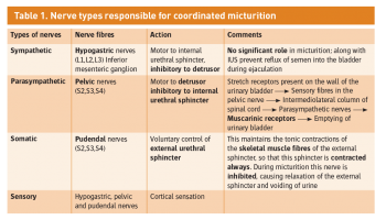
    Table 1. Nerve types responsible for coordinated micturition(click to enlarge)

    Neurological bladder dysfunction

    Cortical bladder

    This is normal in newborns and infants, who have not acquired control of socially acceptable voiding, and therefore leads to periodic complete evacuation. Lesions in the paracentral lobule (cerebral palsy, multiple sclerosis, trauma, infarcts) or advancing Alzheimer’s disease result in uncontrolled evacuation in socially unacceptable situations. Since the pontine arc is intact, evacuation is complete, no residual urine and coordination is good, and there is no detrusor sphincter dyssynergia. The bladder is safe in these circumstances.

    Spinal upper motor neuron lesions: hyper-reflexic bladder

    Spinal cord lesions or transections above the sacral cord and below the pons (transverse myelitis, trauma) will eventually lead to detrusor hyper-reflexia. As with most spinal cord lesions, there is an initial period of spinal shock, which may last from weeks to months. During the acute spinal shock period, the bladder remains atonic with large volumes of urinary retention requiring draining. After this period resolves, bladder tone begins to recover and the bladder once again establishes communication with the sacral spinal cord. A spinal arc is re-established leading to a high pressure, low capacity bladder with empties reflexively through the sacral micturition centre (automated bladder). There is often residual urine due to incomplete evacuation, and detrusor sphincter dyssynergia, predisposing to vesico-ureteric reflux, infections and impaired renal function. 

    Spinal lower motor neuron lesions: areflexic bladder

    Spinal cord lesions are found at or below the sacral cord which leads to a loss of the spinal reflex arc, the sequelae of which is an areflexic, large capacity bladder with overflow incontinence and a high residual volume predisposing individuals to urinary tract infections (UTIs). Sensation may be intact and these may be painful.

    Sensory denervated bladder

    This is usually seen in diabetic neuropathy, tabes dorsalis or even changes associated with chronic urinary retention. This is not usually seen in children, and is characterised by a complete lack of sensation of bladder filling and the subsequent desire to void, and overflow incontinence. These individuals can often void by straining or pressing on their abdomen to overcome their abdominal leak point pressure (ALPP), however emptying is incomplete.

    Non-neurological bladder voiding dysfunction (functional)

    This consists of essentially functional, abnormal patterns of micturition in the presence of an intact neuronal pathway and without any congenital/anatomical abnormality of the urinary tract. This so-called functional urinary incontinence may be caused by disturbances in the filling (storage) phase, the voiding phase or a combination of both.

    Overactive bladder 

    This is characterised by frequent episodes of urgency, frequency and nocturia which in both childhood and adulthood is often countered by contractions of pelvic floor muscles and holding manoeuvres (squatting, crossing legs, sitting on heels). Symptoms are due to underlying detrusor overactivity (filling phase defect, characterised by cystometrography). The bladder capacity is often small, but the voiding pattern is normal with appropriate relaxation of pelvic floor muscles. Constipation can sometimes trigger detrusor contraction by stimulation of stretch receptors in bladder wall by extrinsic faecal mass or by colonic contractions via shared neural pathways.

    Fractionated voiding

    Micturition occurs in several small fractions. Bladder emptying is incomplete due to underactivity of detrusor muscles. Abdominal muscles are then used to increase pressure on the bladder (valsalva voiding). There exists an irregular but continuous flow rate. This may lead to an underactive bladder, which is the long-term result of dysfunctional voiding with detrusor decompensation. There is no longer any detrusor contraction during voiding. A large post residual volume may exist with a predisposition to recurrent UTIs.

    Hinman syndrome 

    This is a rare but serious case of voiding dysfunction, also known as non-neurogenic neurogenic bladder. It is associated with severe bladder sphincter dyssynergia, and a trabeculated bladder, which develops a high-pressure state with bilateral vesicoureteric reflux and a large residual volume. This is akin to a neurogenic bladder, however there is no obvious neurological abnormality. The condition has also been known to lead to renal failure. The syndrome is probably caused by acquired behavioural and psychological disorders manifested by bladder dysfunction mimicking neurologic disease. The dysfunction is associated with abnormal family dynamics in 50% of cases. Individuals under psychosocial pressure try to inhibit enuresis by voluntarily contracting the external sphincter. These voluntary contractions lead to an obstruction of the urinary tract, characterised by an intermittent stream, increased residual urine and increased intravesicular pressure. The resultant destruction of the urinary tract simulates true neurogenic bladder.

    Dysfunctional elimination syndrome

    The co-existence of any of these conditions along with constipation gives rise to the term dysfunctional elimination syndrome. Other dysfunctional voiding patterns not dealt with here include voiding postponement, giggle incontinence, vesico-vaginal entrapment and pollakiuria.

    References

    1. Danforth TL, Ginsberg DA. Neurogenic lower urinary tract dysfunction: how, when, and with which patients do we use urodynamics? Urol Clin North Am 2014 Aug; 41(3):445-52
    2. Unger CA, Tunitsky-Bitton E, Muffly T, Barber MD. Neuroanatomy, neurophysiology, and dysfunction of the female lower urinary tract: a review. Female Pelvic Med Reconstr Surg 2014 Mar-Apr; 20(2):65-75
    3. Gray M. Traces: making sense of urodynamics testing – part 13: pediatric urodynamics. Urol Nurs 2012 Sep-Oct; 32(5):251-5, 274
    4. Woolf AS, Stuart HM, Newman WG. Genetics of human congenital urinary bladder disease. Pediatr Nephrol 2014 Mar; 29(3):353-60. doi: 10.1007/s00467-013-2472-1
    5. Bauer RM, Huebner W. Gender differences in bladder control: from babies to elderly. World J Urol 2013 Oct; 31(5):1081-5. doi: 10.1007/s00345-013-1132-1. Epub 2013 Jul 24
    6. Haifler M, Stav K. Dysfunctional voiding in adults. Isr Med Assoc J 2013 May; 15(5):247-51
    © Medmedia Publications/Hospital Doctor of Ireland 2015